TISSUE
CELL
PATHOLOGY
ANTIBODY INFORMATION
Antibody HPA011271
Antibody HPA011272
Antibody CAB013023
Antibody CAB035987
Antibody CAB058693
Provider
Product name
Host species
Clonality
Purity
Other gene match
Released in version
References
Proper citation
VALIDATION SUMMARY
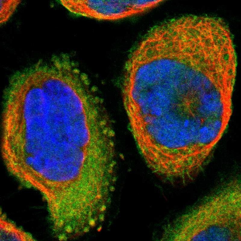
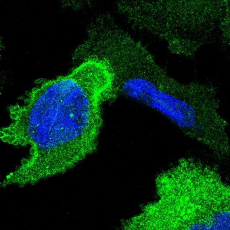
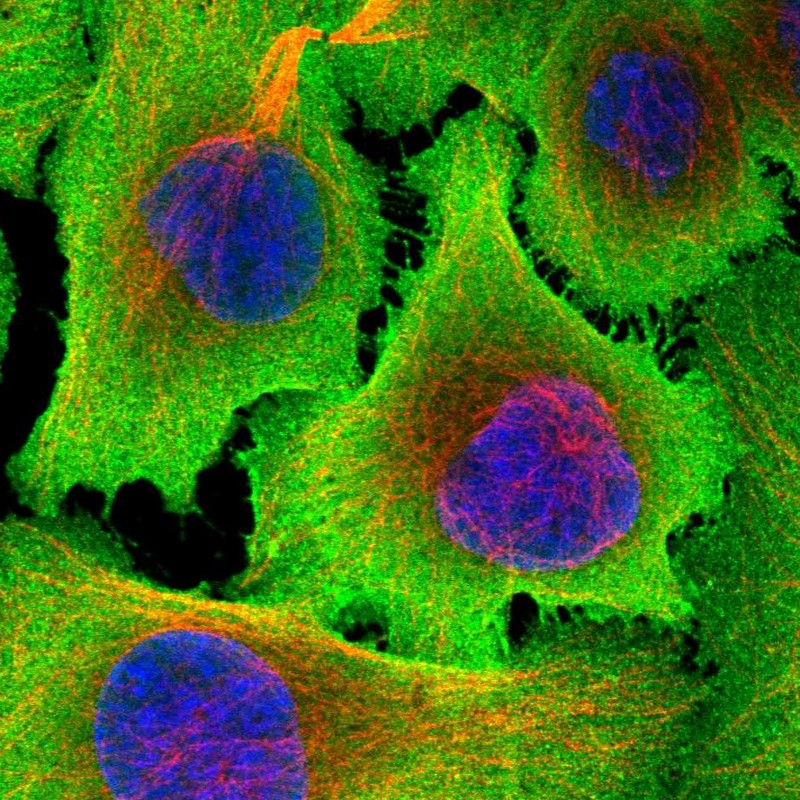
IMMUNOCYTOCHEMISTRY
Formal validation: Independent
Standard validation
Figure description
Antibody dilution
Literature conformity
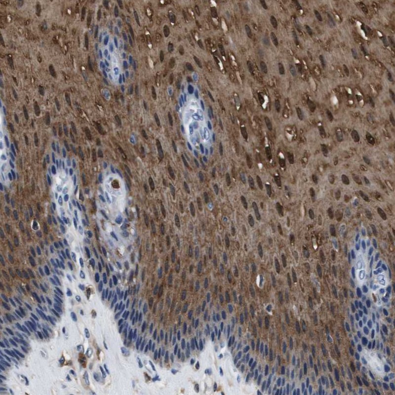
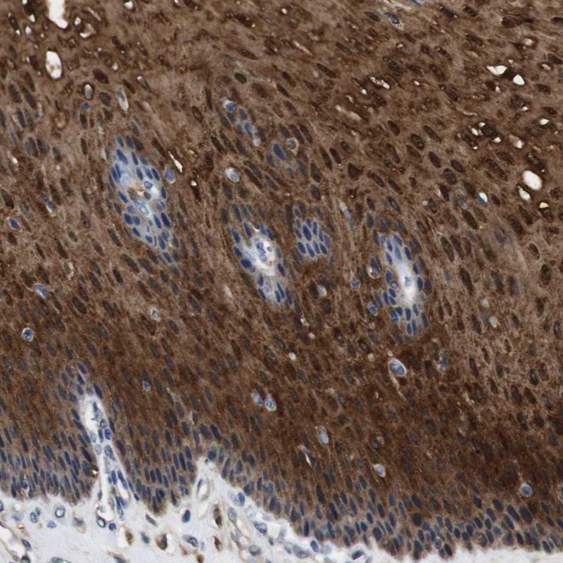
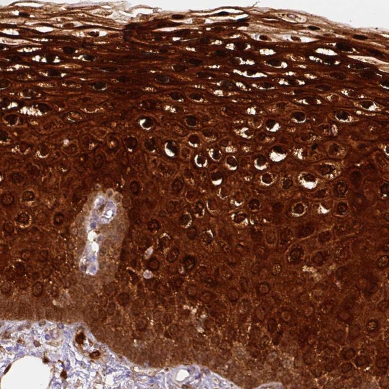
IMMUNOHISTOCHEMISTRY
Formal validation: Orthogonal
Expression
Retrieval
RNA consistency
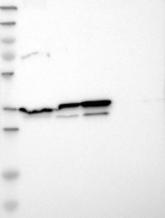
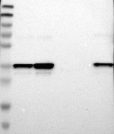
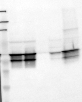
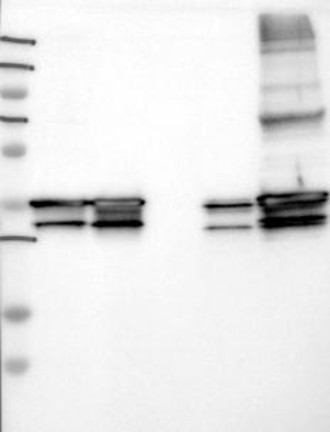
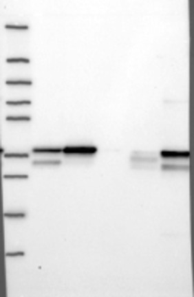
WESTERN BLOT
Target mass (kDa)
PROTEIN ARRAY
ANTIGEN INFORMATION
Antigen
Length (aa)
Antigen sequence
FRNALLSLAKGDRSEDFGVNEDLADSDARALYEAGERRKGTDVNVFNTIL
TTRSYPQLRRVFQKYTKYSKHDMNKVLDLELKGDIEKCLTAIVKCATSKP
AFFAEKLHQAMKGVGTRHKALIRIMV
MAMVSEFLKQAWFIENEEQEYVQTVKSSKGGPGSAVSPYPTFNPSSDVAA
LHKAIMVKGVDEATIIDILTKRNNAQRQQIKAAYLQETGKPLDETLKKAL
TG
Matching transcripts
RELEVANT PUBLICATIONS
1.