TISSUE
CELL
PATHOLOGY
ANTIBODY INFORMATION
Antibody HPA051476
Antibody HPA054698
Antibody CAB016780
Antibody CAB018650
Provider
Product name
Host species
Clonality
Purity
Other gene match
Released in version
References
Proper citation
VALIDATION SUMMARY
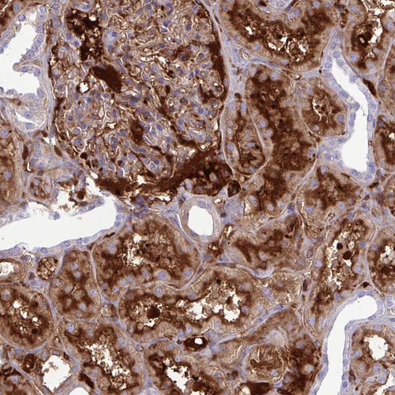
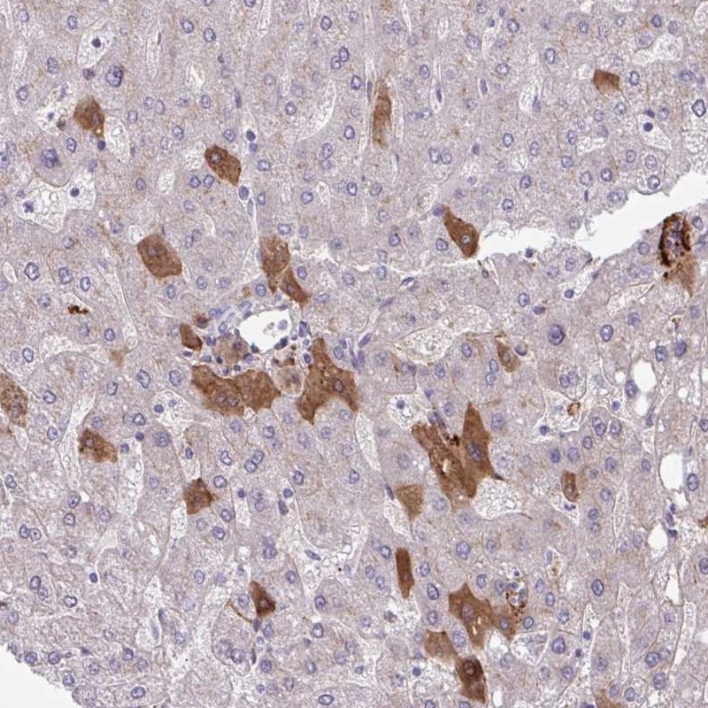
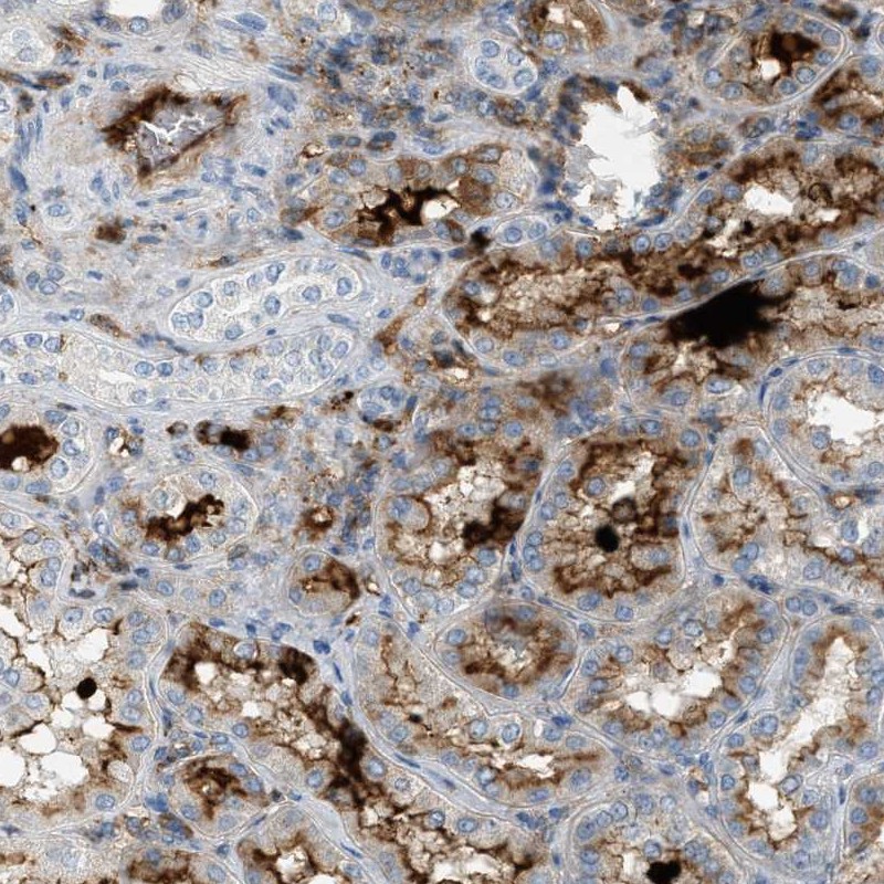
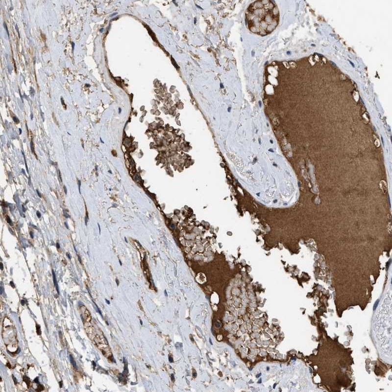
IMMUNOHISTOCHEMISTRY
Formal validation: Orthogonal
Figure description
Formal validation: Independent
Standard validation
Expression
Retrieval
Antibody dilution
Literature conformity
RNA consistency
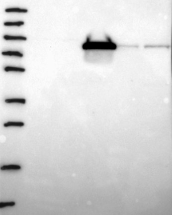
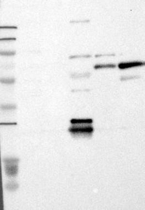
WESTERN BLOT
Target mass (kDa)
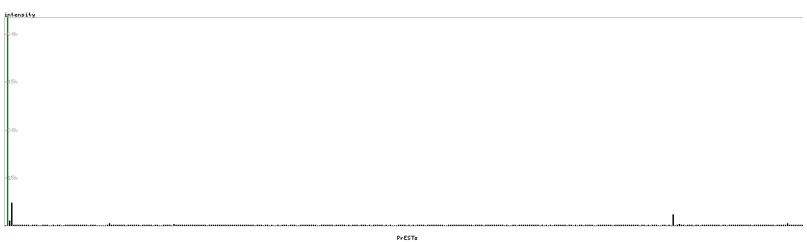
PROTEIN ARRAY
ANTIGEN INFORMATION
Antigen
Length (aa)
Antigen sequence
FEALESSTATDVFWAKYTACETARTPRDKLAACLEGNCAEGLGTNYRGHV
NITRSGIECQLWRSRYPHKPEINSTTHPGADLQ
EGVWCYVAGKPGDFGYCDLNYCEEAVEEETGDGLDEDSDRAIEGRTATSE
YQTFFNPRTFGSGEADCGLRPLFEKKSLE
Matching transcripts