We use cookies to enhance the usability of our website. If you continue, we'll assume that you are happy to receive all cookies. More information. Don't show this again.
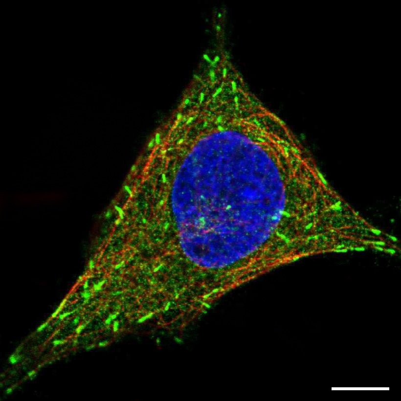
Staining of microtubule end in human cell line SiHa (HPA026678)
Scale bar represents 10µm
Microtubules
Microtubules are a part of the cellular cytoskeleton and function as a rail network within the cytoplasm used to transport vesicles to different locations. Their polymerization starts from the microtubule-organizing center (MTOC) and stretches out to the edges of the cell. A characteristic feature of microtubules is that they are constantly in the process of elongation (polymerization) or shrinkage (depolymerization), a phenomenon known as dynamic instability. This allows the cell to change its shape in response to different environmental conditions.
Microtubule ends
The process of dynamic instability is much faster at the free ends of the microtubules, thus referred to as the plus ends, whereas the slowly changing minus ends are joined together at the MTOC.
Mitotic spindle
During mitosis the microtubule network will disassemble completely and instead form the mitotic spindle, which is responsible for separating the duplicated chromosomes into the two daughter cells.
Immunofluorescent staining
Immunofluorescent staining of microtubules shows thin strands that stretch throughout the whole cell. It is almost always possible to detect the site from which they originate, the MTOC. Some proteins are exclusively localized to the growing plus ends of the microtubule, in which case only the tip furthest away from the center is stained. The mitotic spindle can be detected during cell division as a structure that attaches to the chromosomes and pulls them apart in the process. Size and shape of the mitotic spindle vary and are dependent on the progress of the cell division.