We use cookies to enhance the usability of our website. If you continue, we'll assume that you are happy to receive all cookies. More information. Don't show this again.
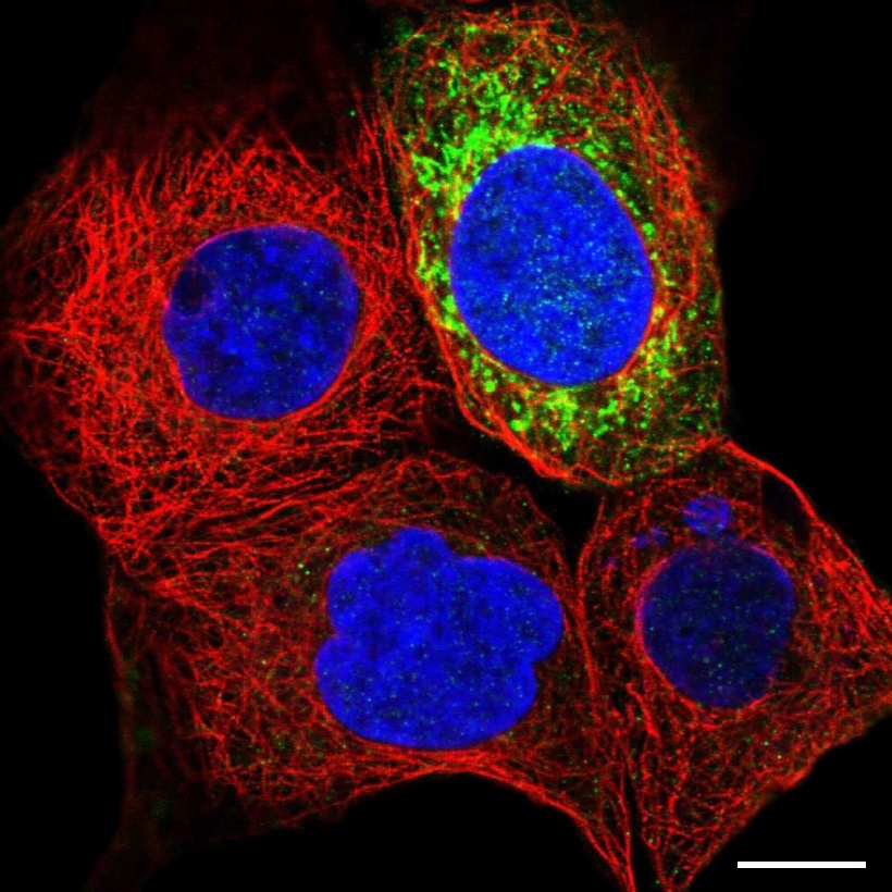
Staining of scv intensity in human cell line A-431 (HPA022522)
Scale bar represents 10µm
Single-cell variation
As the images in the Cell Atlas provide single cell resolution, variations in protein expression patterns from cell to cell can be observed. Since the cells are grown under asynchronous conditions the most likely explanation for this would be cell cycle effects. Other factors could for example be environmental conditions, cell confluency, cellular stress or stochasticity.
Immunofluorescent staining
In our confocal images both variation in the abundance (SCV intensity) or a spatial re-distribution (SCV spatial) of the target protein, can be revealed. Confocal microscopy enables the acquisition of an image in a narrow optical section. When observing a cell-to-cell variation pattern in an IF image it is important to ensure that the optical section is consistent for all cells in the image by taking caution to the reference markers.
SCV intensity A single-cell variation (intensity) is defined as intensity differences of the immunostaining between cells. This variance in staining intensity reflects different levels of expression of the target protein.
SCV spatial A single-cell variation (spatial) is characterized by immunostaining of different cellular compartments in different cells. This could indicate a dynamic protein expression pattern and potential translocations between respective compartments.