We use cookies to enhance the usability of our website. If you continue, we'll assume that you are happy to receive all cookies. More information. Don't show this again.
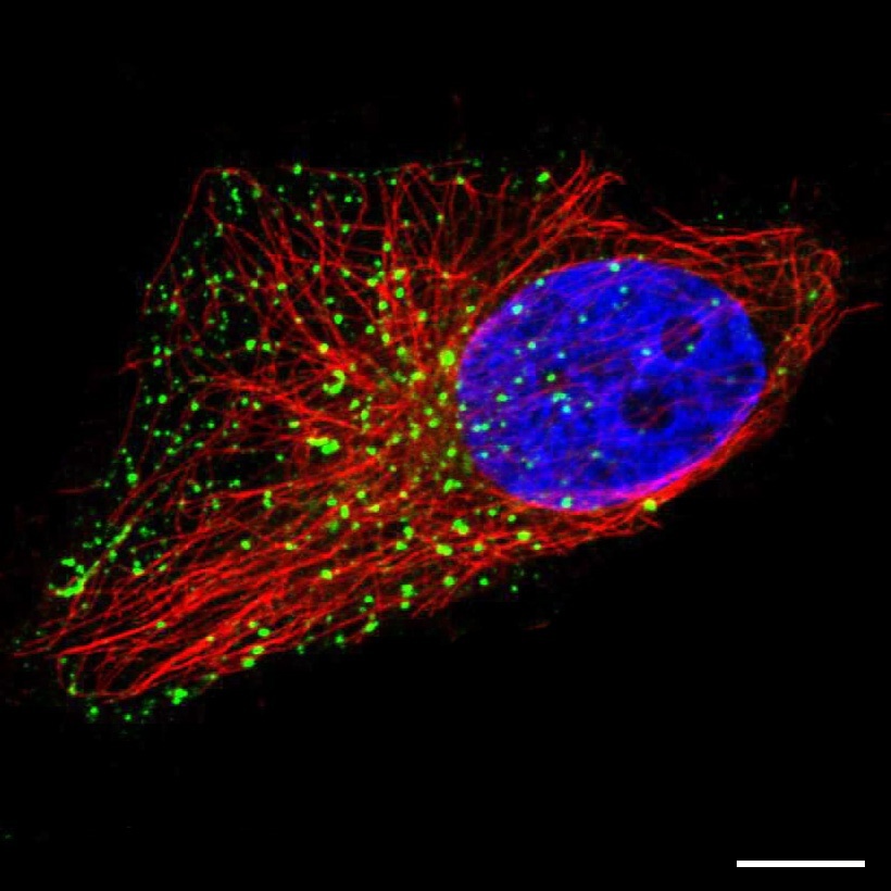
Vesicle is a collective term for a number of different, small membrane-bound organelles. They are often involved in the intracellular transport of proteins and membranes between organelles and the release of substances to the outside of the cells (secretory vesicles), or have specialized metabolic functions. Four examples of vesicle-like organelles that are commonly found in the human cell are listed below.
Endosomes
Endosomes are the main organelles for sorting material that has been taken up from the exterior of the cell by the plasma membrane in a process called endocytosis. The endosomes can then either recycle its contents by bringing it back to the plasma membrane or degrade it by fusing with a lysosome.
Lysosomes
Lysosomes are filled with hydrolytic enzymes and are responsible for degrading molecules within the cell. They can fuse with late endosomes, creating so-called endolysosomes, which degrade the material within the endosome.
Peroxisomes
Peroxisomes contain enzymes that are linked to various metabolic pathways. They are mainly involved in the breakdown of fatty acids but also necessary for the degradation of hydrogen peroxide.
Lipid droplets
Lipid Droplets are specialized organelles for the cellular storage of neutral lipids. They vary in their size and have a unique structure consisting of a hydrophobic core containing the lipids, and a surrounding phospholipid monolayer with attached proteins.
Immunofluorescent staining
Vesicle stainings usually appear as small and bright dots in the cytoplasm but can vary a lot in number, size and distribution throughout the cell. Stainings of larger lipid droplets can be identified, as they appear in a round, ring-like pattern. It is much more difficult to distinguish between the other kind of vesicles solely based on the staining pattern: endosomes are usually located closer to the nucleus and the Golgi apparatus. Lysosomes are also often located closer to the Golgi apparatus, but they are larger and more densely stained. Peroxisomes are spread throughout the cell and possess a more elongated shape.
Although the staining pattern can indicate towards a type of vesicle, there is no certainty without further validation by other methods like co-localization with typical marker proteins or dyes. Therefore vesicle-like stainings are annotated as vesicles in the Human Protein Atlas and the terms "endosomes", "lysosomes" and "peroxisomes" are only used if co-localization experiments were carried out.