Bronchus
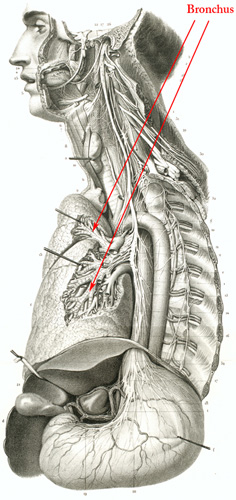
Bronchus
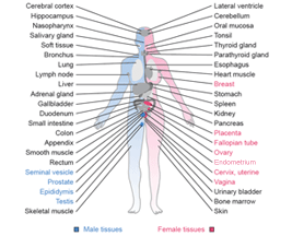
Bronchi are tube like structures that allow air to be transported from the surrounding atmosphere to the lung. The respiratory system (airways) consists of the trachea (essentially a large bronchi) that branch off into smaller and smaller bronchi before reaching the most distal lung alveoli, where oxygen can be transferred to the blood.
The respiratory bronchus is lined by respiratory epithelium comprising an admixture of pseudostratified ciliated columnar epithelium, goblet cells (mucin producing cells also termed mucous cells) and basal cells that function as progenitor cells for both the ciliated columnar cells and goblet cells. Ciliated cells are about five times more numerous than goblet cells in the central airways and the ratio of ciliated cells increases in the smaller and peripheral bronchi (bronchioles) as the goblet cells diminish. The basal cells are more abundant in the proximal airways, where they form more or less, a continuous layer and gradually diminish in number in peripheral bronchi. Small clusters of neuroendocrine cells located within the bronchial epithelium called neuroepithelial bodies can be identified.
The subepithelial tissue can be divided into a lamina propria situated between the basement membrane and the muscularis mucosae, and a submucosa. The lamina propria contains a network of capillaries, reticulin fibers and bundles of elastic and nerve fibers. The submucosa of larger bronchi contains cartilage with chondrocytes, muscle tissue as well as serous and mucous glands, termed peribronchial glands. Anatomically, three regions can be recognized in these glands: the ciliated duct, the connecting duct, and the secretory tubules lined by serous and mucinous glandular cells. A myoepithelial cell layer is also present.
Cancer: Lung cancer
|