We use cookies to enhance the usability of our website. If you continue, we'll assume that you are happy to receive all cookies. More information. Don't show this again.
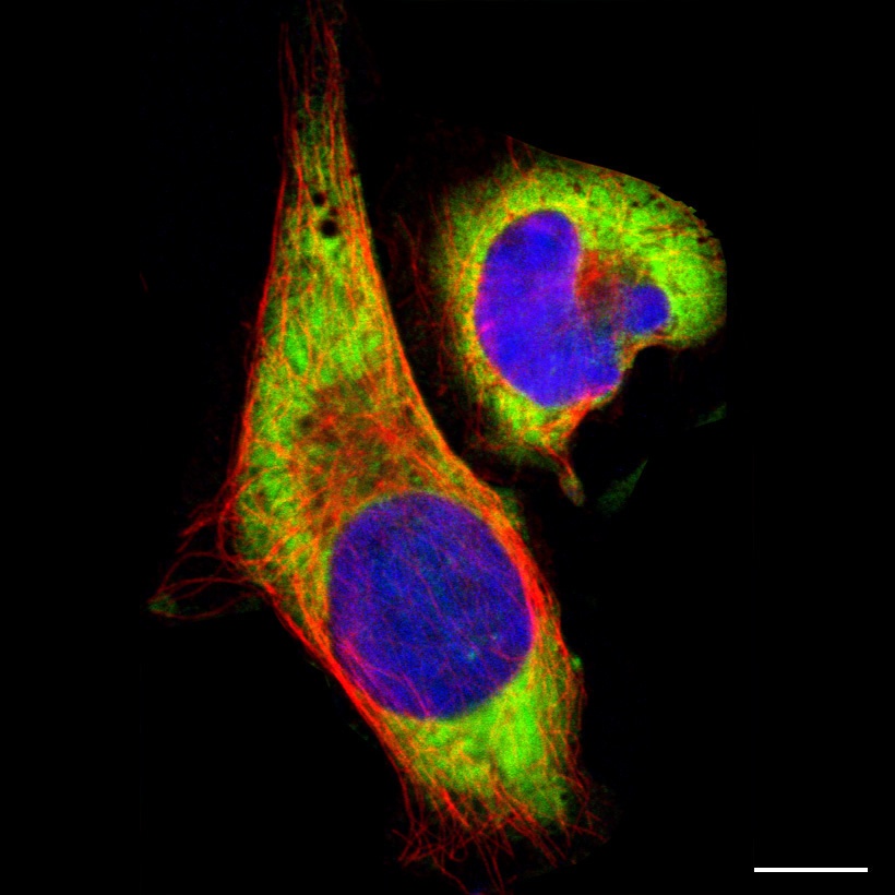
Staining of cytoplasm in human cell line U-2 OS (HPA003570)
Scale bar represents 10µm
Cytosol
The cytosol is referred to as the part of the cytoplasm that is not a membrane bound organelle or a cytoskeleton. It is a liquid matrix embedding the organelles, making up about 70% of the cell volume and is mainly composed of water. The cytosol plays an important role in the cell homeostasis as the site of many metabolic reactions and in the storage of free proteins as well as ions such as potassium, sodium and bicarbonate.
Immunofluorescent staining
The immunofluorescent staining of the cytoplasm encompasses the whole cell but the nucleus. The characteristic of the staining can vary from smooth to granular and is often stronger closer to the nucleus.