We use cookies to enhance the usability of our website. If you continue, we'll assume that you are happy to receive all cookies. More information. Don't show this again.
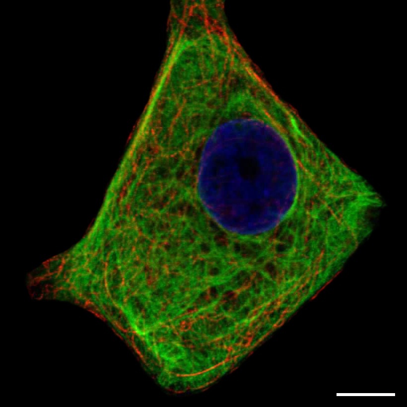
Staining of intermediate filaments in human cell line MCF-7 (HPA002465)
Scale bar represents 10µm
Intermediate filaments
Intermediate filaments are a part of the cellular cytoskeleton. They have a coiled structure that provides mechanical support to the cell as they stretch from the nucleus throughout the cytoplasm. Cells that are subject to mechanical stress, such as hair and skin cells, contain a higher amount of intermediate filaments compared to other cell types. Additionally intermediate filaments participate in the organization of chromatin in the nucleus. The latter is achieved by anchoring the chromatin to the nuclear lamina that lines the inner part of the nuclear membrane.
Immunofluorescent staining
Immunofluorescent staining of intermediate filaments can vary a lot between cell types. The filaments are usually at least partly located close to the nucleus, but may stretch throughout the whole cell. Common for most stainings is that the filaments exhibit a tangled structure with strands crossing every so often. Sometimes the staining can be mistaken for microtubules at first, but there is little or no overlap when comparing with the microtubule marker (red channel).