We use cookies to enhance the usability of our website. If you continue, we'll assume that you are happy to receive all cookies. More information. Don't show this again.
Antibody staining overlaps with antibody HPA038435.
Validated
Antibody staining overlaps with antibody HPA038434.
Standard validation
Supported
Supported
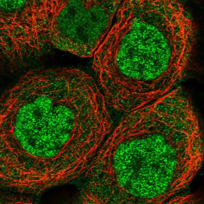
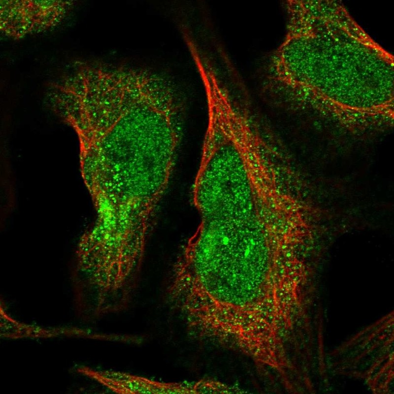
Figure description
Immunofluorescent staining of human cell line A-431 shows localization to nucleoplasm & cytosol.
Immunofluorescent staining of human cell line U-2 OS shows localization to nucleoplasm & cytosol.
Antibody dilution
1:154
1:24
Literature conformity
The subcellular location is supported by literature.
The subcellular location is supported by literature.
WESTERN BLOT
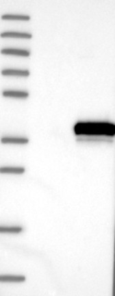
Antibody HPA038434
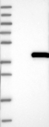
Antibody HPA038435
Standard validation
Supported
Single band corresponding to the predicted size in kDa (+/-20%).
Supported
Single band corresponding to the predicted size in kDa (+/-20%).
Figure description
Lane 1: Marker [kDa] 250, 130, 95, 72, 55, 36, 28, 17, 10 Lane 2: Negative control (vector only transfected HEK293T lysate) Lane 3: Over-expression Lysate (Co-expressed with a C-terminal myc-DDK tag (~3.1 kDa) in mammalian HEK293T cells, LY412885)
Lane 1: Marker [kDa] 250, 130, 95, 72, 55, 36, 28, 17, 10 Lane 2: Negative control (vector only transfected HEK293T lysate) Lane 3: Over-expression Lysate (Co-expressed with a C-terminal myc-DDK tag (~3.1 kDa) in mammalian HEK293T cells, LY412885)
Target mass (kDa)
39.1, 38.2, 38.1
39.1, 38.2, 38.1
Antibody dilution
1:250
1:250
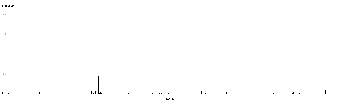
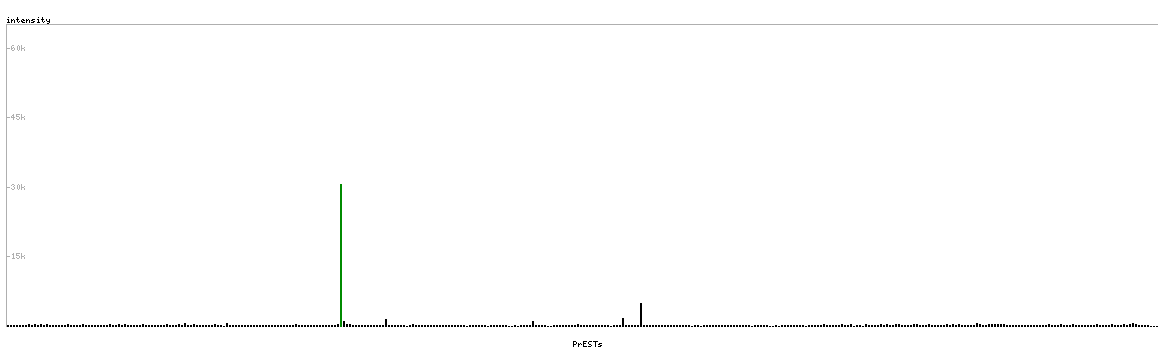
PROTEIN ARRAY
Antibody HPA038434
Antibody HPA038435
Standard validation
Approved
Pass with quality comment low specificity (binding to 1-2 PrESTs >15% and <40%).
Approved
Pass with quality comment low specificity (binding to 1-2 PrESTs >15% and <40%).
Figure description
Antibody specificity analysis with protein arrays. Predicted and matching interactions are shown in green.
Antibody specificity analysis with protein arrays. Predicted and matching interactions are shown in green.