Colorectal cancer
Male, 77 years, moderately differentiated adenocarcinoma, Stage I (T1, N0, M0), adenomatous colon mucosa left lower part of image 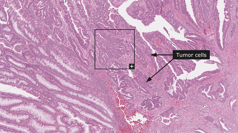
Colorectal cancer
Colorectal cancer (CRC) is the second most common cancer in the industrialized world, accounting for approximately one million new cases each year. Incidence rates correlate with meat consumption and migration studies reinforce the importance of environmental factors. The overall mortality is approximately 50%. The surgical stage at diagnosis is the most important factor for predicting patient outcome, with five year survival rates of more than 90% for stage I disease and less than 10% for stage IV disease.
The surgical stage represents a classification system based on the extent and depth of tumor growth. Stage I CRC shows invasive grown into the anatomical layers of the colon, but the tumor is not spread outside the colon wall or into regional lymph nodes. Stage II CRC demonstrates extended growth through the outer layer of the colon (peritoneum) and may have extended into nearby organs, but has not spread to any lymph node. Stage III CRC have spread to nearby lymph nodes but not yet metastasized to distant sites in the body. Finally, in Stage IV CRC the tumor has spread to distant organs such as the liver, lungs, or other sites. The Dukes classification is an older and less complicated staging system that predates the TNM system, and translates so that Duke A= Stage I, Duke B= Stage II, Duke C= Stage III and Dukes D= Stage IV.
CRC is considered to develop through a multi-step process, originating from normal colon epithelium that develops into precursor lesions termed adenomas. Adenomas can subsequently further progress into invasive CRC with metastatic potential. Spread of CRC occurs by direct growth through the bowel wall and through invasion of lymphatic and venous channels. The most common sites for metastases are regional lymph nodes and the number of lymph node metastases influences prognosis. The liver is the most common distant site for CRC metastases.
The vast majority of CRC are adenocarcinomas, with less than 10% of the cancers being distinguished by an abundant secretion of mucin. The tumors are classified according to the degree of morphological differentiation into well, moderately and poorly differentiated. About 80% are well or moderately differentiated with a growth pattern consisting of tumor cells that form irregular glandular structures present at different layers of the bowel wall. Poorly differentiated CRC show no, or only hinted, glandular formation. Overall poor differentiation with a diffuse infiltrative growth pattern is associated with poor prognosis, although stringent classification systems based on morphological features are lacking. Treatment decisions are thus based on the surgical stage without consideration to morphological characteristics. In addition to adenocarcinomas, endocrine tumors can also arise within the colorectal mucosa. Squamous and adenosquamous tumors are exceedingly rare.
In addition to the diagnostic procedure, which is based on microscopical examination of a tumor specimen, immunohistochemistry can be used to determine a colorectal origin of a metastasis or to visualize the spread of tumor cells in surrounding tissues. Antibodies that show high sensitivity and specificity for tumors of colorectal origin include Cytokeratin 20, CDX-2, SATB2 and Cadherin-17. Chromogranin-A antibodies can be used to distinguish endocrine tumors in the bowel from common adenocarcinomas.
Recently new targeted drugs have been implemented into the treatment of patients with advanced colorectal cancer. Epidermal growth factor receptor (EGFR) is commonly expressed in colorectal tumors and monoclonal antibodies inhibiting EGFR demonstrate clinical efficacy in patients with tumors that do not harbor downstream activating KRAS mutations. Today KRAS mutation status is analyzed routinely before starting anti-EGFR treatment.
Normal tissue: Colon, Rectum
|