Enzyme-linked immunosorbent assay and microarrays
Since the very first use of antibodies for the detection of antigens, many different technologies have been developed that make use of the antibodies' capability to bind to other molecules.
During the 1950s, the scientists Yalow and Berson developed a method where radioactivity is used to determine the amount of an analyte in a solution. This so called 'radioimmunoassay' (RIA), for which Yarlow received the Nobel prize in 1977, was a very sensitive method for the detection of hormones but using radioactivity for antigen detection is not safe and suitable for a general use. Hence, an alternative procedure was developed by linking enzymes to antibodies instead of a radioactive molecule, and by adhering molecules to surfaces. In one of the nowadays most common applications today are measuring the quantity of a biomolecule in a sample by "enzyme-linked immunosorbent assay" (ELISA). This term originally refers to the use of an enzyme to report an interaction
between an antibody and its binding partner (Gan & Patel, 2013). The foundation for Perlmann and Engvall in Sweden
(Engvall & Perlmann, 1971) as well as Schuurs and van Weemen from the Netherlands
(Van Weemen & Schuurs, 1971), who built assays with immobilized and enzyme-modified reagents in the early 1970s.
Today, scientists also use colored molecules (so called fluorophores) that re-emit light upon excitation to visualize antibody-antigen interactions. Many variants of experimental procedures have been developed,
and it is common to build assays using more than one antibody to detect a target of interest (see Figure 1 C-D).
To further enhance the possibilities offered by the immunoassay format, applications based on microarrays have been developed and which allow to measure more than one molecule in a single reaction chamber (see below).
Technology
Assay designThe use of antibodies allows designing experiments in many different ways for the intended analysis. To achieve the best possible results from the experiment, different reagents, additives, and solutions have to be
tested for their optimal combination and concentration, incubation times and the number of wash cycles need to be evaluated and adjusted. This is to avoid unwanted interactions, which disturb the analysis from
detecting the target of interest. Moreover, the mode of how a target is identified and detection can be performed in a number of ways, as described in Figure 1.
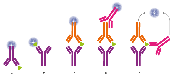
Figure 1. Different setups for ELISA and other immunossays.
In ELISA assays, the antibodies may (A) detect an immobilized antigen, (B) capture a labeled antigen, (C) capture an unlabeled antigen and use a second, labeled antibody to detect the captured antigen, or
(D) use a third antibody for detection, or even use two antibodies for detection (E) (see Proximity Ligation Assay).
Direct labeling of the antibody or antigen as in (A), (B), and (C) is the simplest and fastest method for detection. Using a secondary antibody as detection method, as shown in (D) and (E), will further increase
the sensitivity and selectivity of the analysis. The method used in (D) also allows greater flexibility, whereas method (E) further increases the specificity, as three antibodies must bind the antigen in order to
produce a reporter molecule. Out of the presented assays, the most commonly used concepts are shown in (C) and (D).
MultiplexingA new era in immunoassays started with the development of a technology called microarrays. The term microarray most commonly describes the ordered organization of small volume droplets that have dried on a small surface area. The reaction dimensions are miniaturized so that many assays can be performed in multiple samples in parallel, several thousands of different features may be presented to the surrounding solution. This means that scientists can measure a large number of molecules with one single experiment. There is the possibility to use microscope glass slides and specialized robotics that deposit very small drops of liquid
(1 nl = 0.000000001 liter) on the glass surface in an ordered fashion. This leaves behind spots of less than one millimeter in diameter (0.15 mm). Another common technique for multiplexing is to use even smaller and color-coded particles (diameter of 0.005 mm). These particles can be coated with antibodies to fish out the analyte from the solution.
SensitivityIn many applications it is important to measure very small amounts (sometimes only traces) of a molecule in a given sample. In order to achieve the required sensitivity, the conditions of the experiment need to be adjusted to suit the antibodies,
the detection system, and the type of samples. In addition, there is progress being made on using better colors,signal amplification specialized lasers and filters, as well as miniaturization (Ekins & Edwards, 1997).
Specific examples
There are many examples of how ELISA assays may be used in basic research and in clinical diagnostics. One specific example is the sensitive sandwich-type enzyme-linked immunoassay used to determine the amount of the
protein prostate-specific antigen (PSA), which is a biomarker used to detect prostate cancer (Kuriyama et al., 1980).
Microarray assays on the other hand, have previously received a lot of attention for their use in parallel analysis of DNA and RNA molecules. To translate their advantages to assays for the analysis of proteins with antibodies,
new protocols and routines had to be developed and established. Nowadays, there are multiplexed techniques for measuring the amount of proteins in different sample types (e.g. cells, blood serum, urine), to determine how
proteins are modified in biological processes (e.g. phosphorylation), or to describe specific protein-protein interactions. Another example is the analysis of antibodies circulating in blood from patients.
Microarray-based applications have also been built for purified antibodies and to study the antibody binding characteristics an important aspect when using binding reagents as research reagents. Such protein
microarrays can either consist of proteins, protein fragments, or small peptides to test the specificity of the binding reagent. Protein microarrays can reveal the interactions to entire proteins or larger
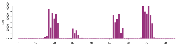
protein fragments, while peptide microarrays show to which particular parts (epitopes) of the proteins the antibodies bind. A typical epitope mapping result is shown in Figure 2
(Edfors et al., 2014). Synthesizing millions of overlapping peptides with only one amino acid residue shift on such arrays enables the mapping of antibody binding regions at
high resolution. This gives very detailed information of the linear (continuous) epitopes recognized by an antibody. Just like with proteins, protein fragments or other antigens, the assembly of peptides on arrays
may also be used for studies of antibody reactivity in plasma samples from patients with infectious and autoimmune diseases.
Figure 2. Epitope mapping of a polyclonal antibody on a peptide array where the result displays four distinct linear epitopes and the consecutive overlapping peptides which are bound. X-axis: peptides, Y-axis: mean fluorescence intensity (MFI). (Edfors et al., 2014)
References and Links
- Edfors, F., Boström, T., Forsström, B., Zeiler, M., Johansson, H., Lundberg, E., ... Uhlen, M. (2014). Immuno-proteomics using polyclonal antibodies and stable isotope labeled affinity-purified recombinant proteins. Molecular & Cellular Proteomics: MCP, 13(6), 1611-1624.
DOI:10.1074/mcp.M113.034140. PubMed: 24722731
- Ekins, R., & Edwards, P. (1997). On the meaning of "sensitivity". Clinical Chemistry, 43(10), 1824-31.
PubMed: 9341999
- Engvall, E., & Perlmann, P. (1971). Enzyme-linked immunosorbent assay (ELISA). Quantitative assay of immunoglobulin G. Immunochemistry, 8(9), 871-4.
PubMed: 5135623
- Gan, S. D., & Patel, K. R. (2013). Enzyme immunoassay and enzyme-linked immunosorbent assay. The Journal of Investigative Dermatology, 133(9), e12.
DOI:10.1038/jid.2013.287. PubMed: 23949770
- Kuriyama, M., Wang, M. C., Papsidero, L. D., Killian, C. S., Shimano, T., Valenzuela, L., ... Chu, T. M. (1980). Quantitation of prostate-specific antigen in serum by a sensitive enzyme immunoassay. Cancer Research, 40(12), 4658-62.
PubMed: 6159971
- The Nobel Prize in Physiology or Medicine 1977. (n.d.). Retrieved February 21, 2014, from http://www.nobelprize.org/nobel_prizes/medicine/laureates/1977/
- Van Weemen, B. K., & Schuurs, A. H. W. M. (1971). Immunoassay using antigen-enzyme conjugates. FEBS Letters, 15(3), 232-236.
PubMed: 11945853
- ELISA, a common technique that uses an enzyme as reporter:
http://en.wikipedia.org/wiki/ELISA
- Eva Engwall, one of the scientists who invented ELISA in 1971:
http://en.wikipedia.org/wiki/Eva_Engvall
- A flat plate with multiple "wells" used as small test tubes, commonly used to perform an ELISA:
http://en.wikipedia.org/wiki/Microtiter_plate
- Immunoassay format may be miniaturized on microarrays to allow multiplexing for multi-parameter analysis:
http://en.wikipedia.org/wiki/Microarray
http://en.wikipedia.org/wiki/DNA_microarray
http://en.wikipedia.org/wiki/Protein_microarray
- Antibodypedia - An open-access database of publicly available antibodies and their usefulness in various applications:
http://www.antibodypedia.com
|