TISSUE
CELL
PATHOLOGY
ANTIBODY INFORMATION
Antibody HPA035248
Antibody HPA057936
Antibody CAB033218
Antibody CAB062556
Provider
Product name
Host species
Clonality
Purity
Other gene match
Released in version
References
Proper citation
VALIDATION SUMMARY
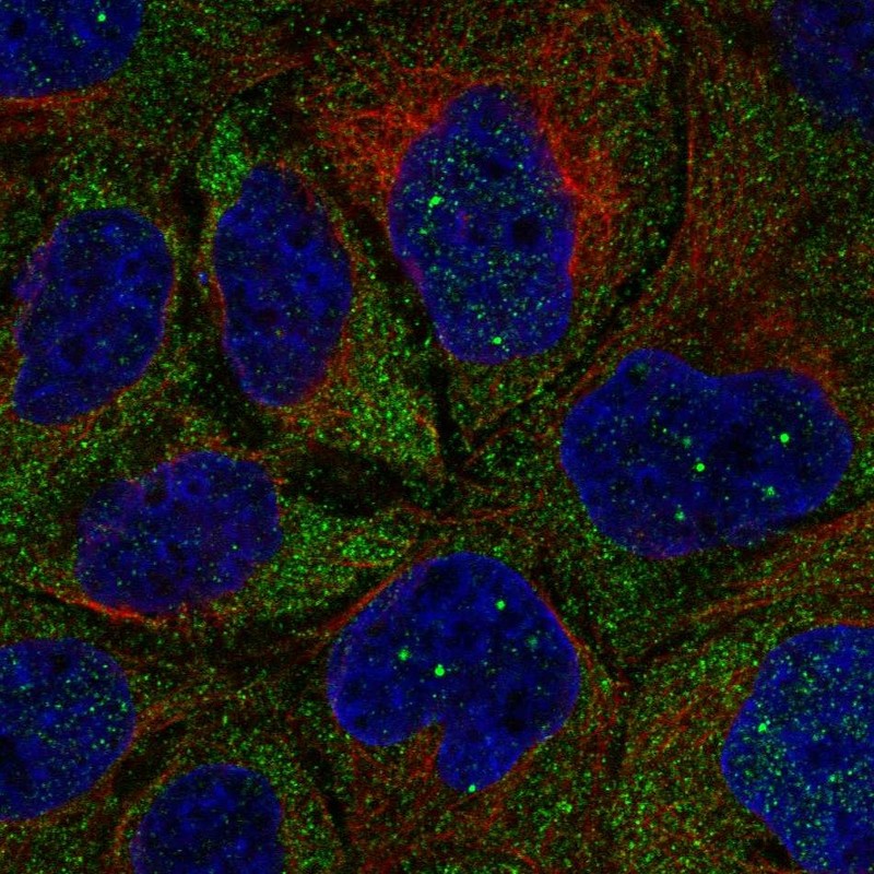
IMMUNOCYTOCHEMISTRY
Standard validation
Figure description
Antibody dilution
Literature conformity
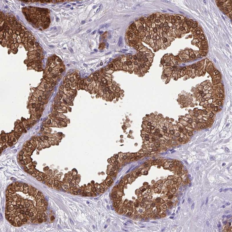
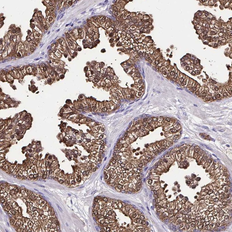
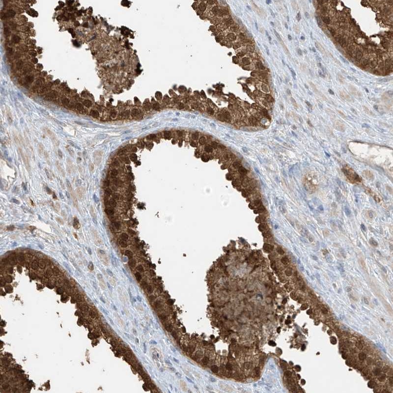
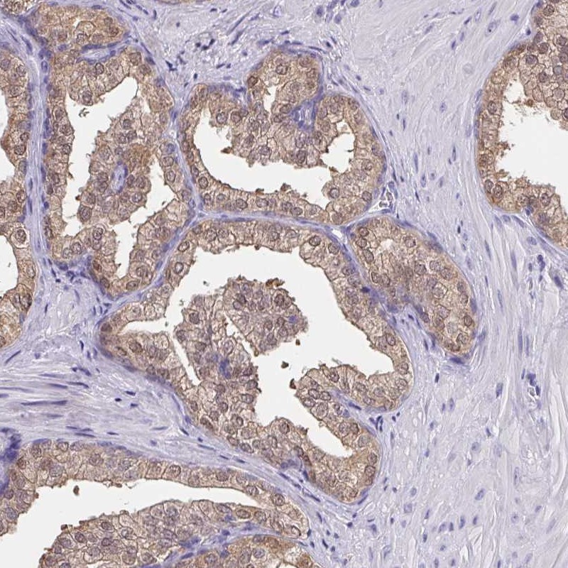
IMMUNOHISTOCHEMISTRY
Formal validation: Orthogonal
Formal validation: Independent
Expression
Retrieval
RNA consistency
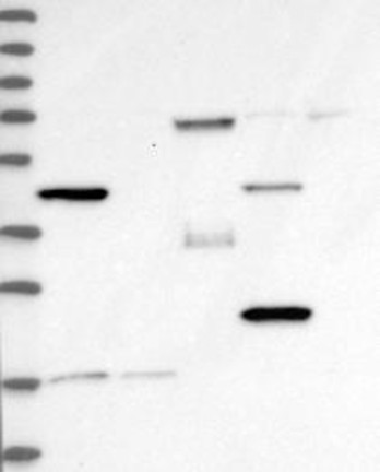
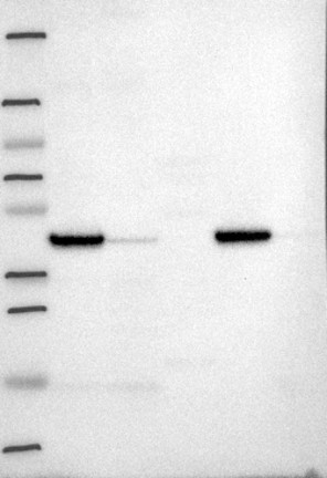
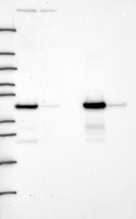
WESTERN BLOT
Target mass (kDa)
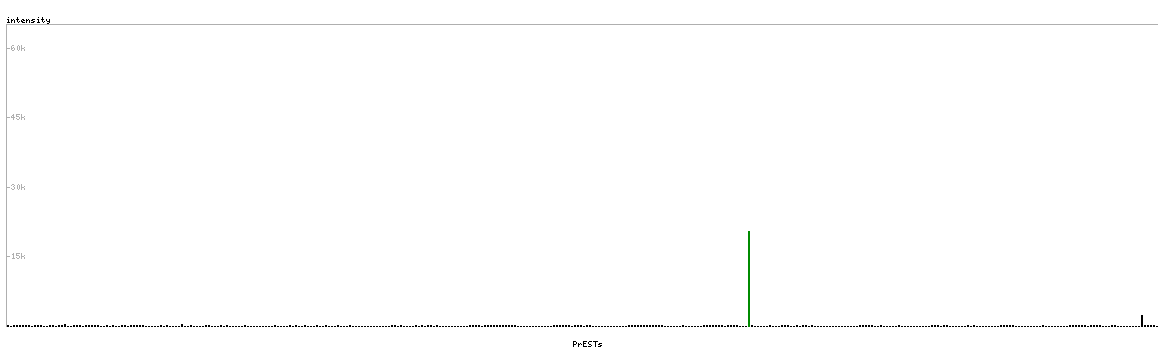
PROTEIN ARRAY
ANTIGEN INFORMATION
Antigen
Length (aa)
Antigen sequence
FVVPGPGKVEITYTPSDGTQKVTYLVHNFEEGGGVAMGMYNQDKSIEDFA
HSSFQMALSKGWPLY
LAHRAKLDNNKELAFFANALEEVSIETIEAGFMTKDLAACIKGLPNVQRS
DYLNTFEFMDKLGENLKIKLAQA
Matching transcripts
RELEVANT PUBLICATIONS
1.