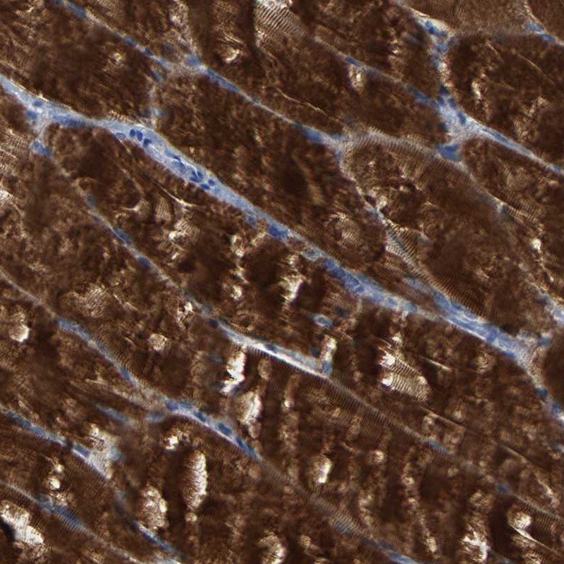
TISSUE
CELL
PATHOLOGY
ANTIBODY INFORMATION
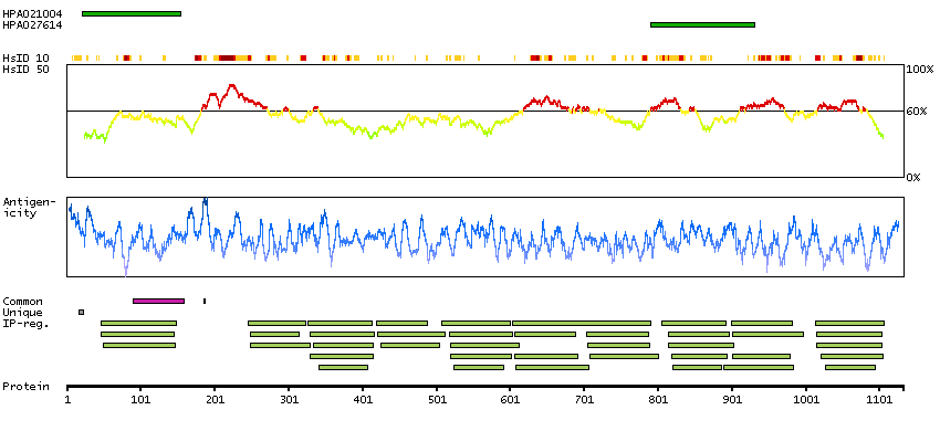
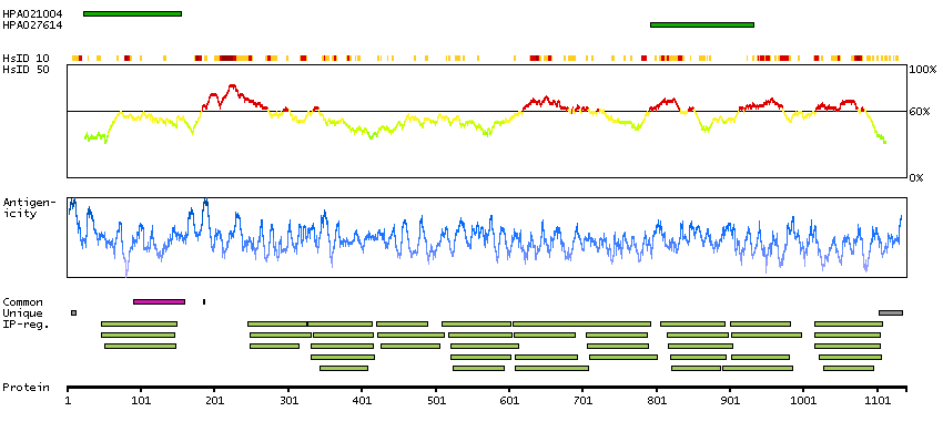
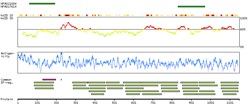
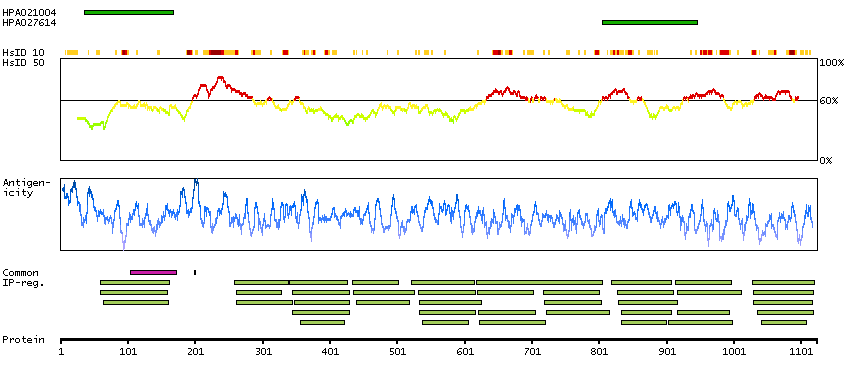
Antibody HPA021004
Antibody HPA027614
Provider
Product name
Host species
Clonality
Purity
Other gene match
Released in version
References
Proper citation
VALIDATION SUMMARY
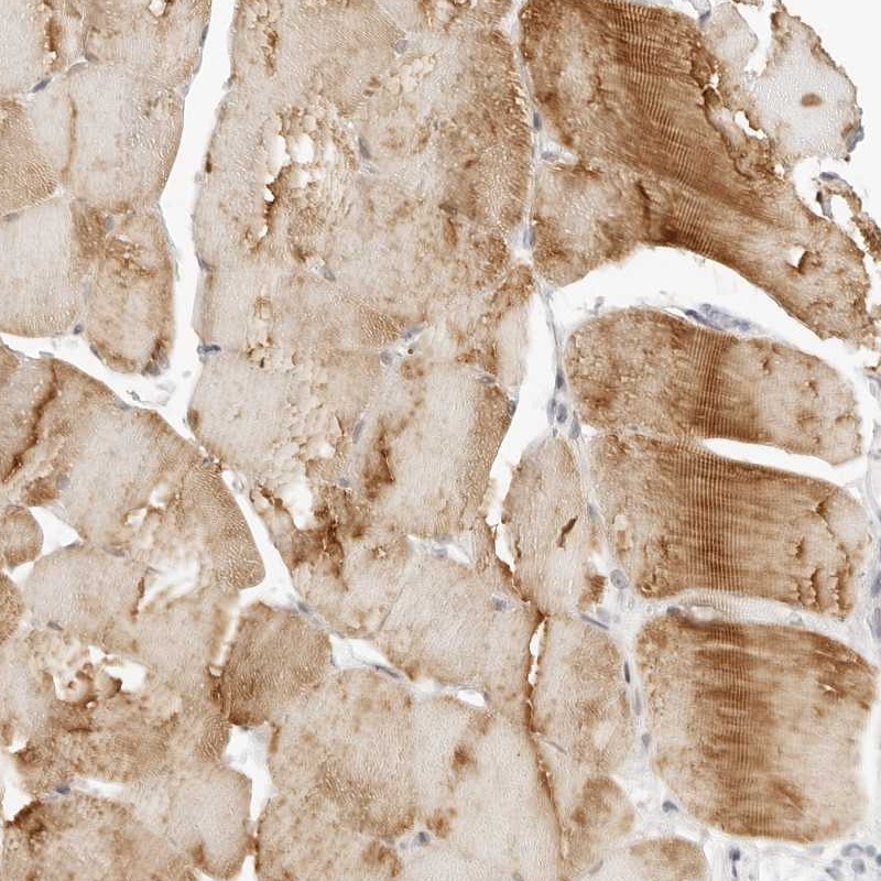
IMMUNOHISTOCHEMISTRY
Standard validation
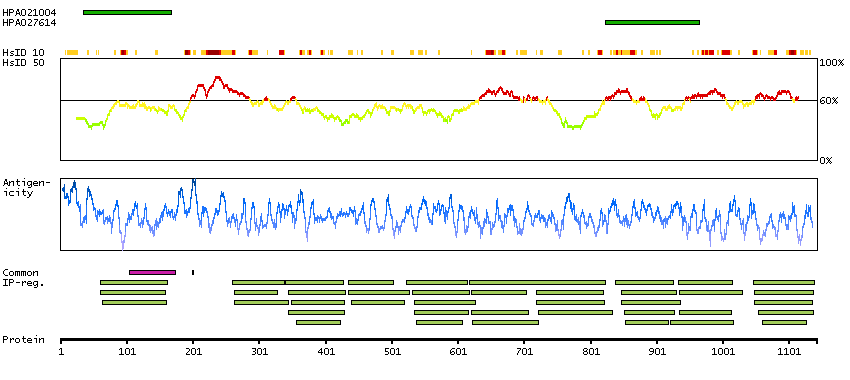
Figure description
Expression
Retrieval
Antibody dilution
Literature conformity
RNA consistency
WESTERN BLOT
Target mass (kDa)
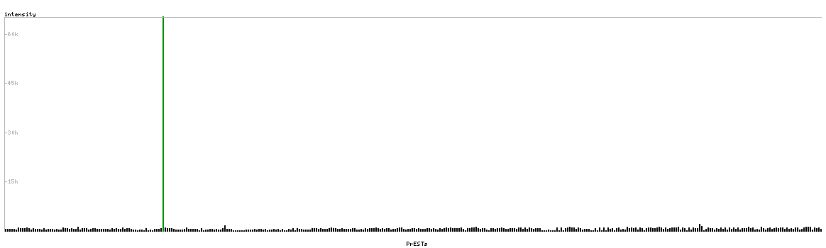
PROTEIN ARRAY
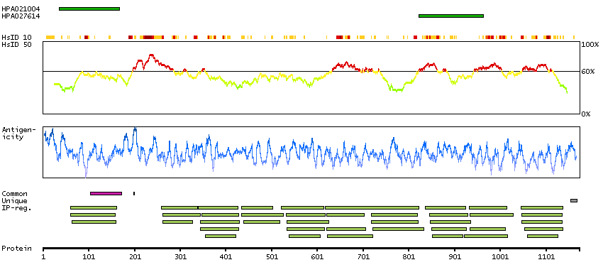
ANTIGEN INFORMATION
Antigen
Length (aa)
Antigen sequence
DWTLVETPPGEEQAKQNANSQLSILFIEKPQGGTVKVGEDITFIAKVKAE
DLLRKPTIKWFKGKWMDLASKAGKHLQLKETFERHSRVYTFEMQIIKAKD
NFAGNYRCEVTYKDKFDSCSFDLEVHESTGTTPN
YYSQPILVKEIIEPPKIRIPRHLKQTYIRRVGEAVNLVIPFQGKPRPELT
WKKDGAEIDKNQINIRNSETDTIIFIRKAERSHSGKYDLQVKVDKFVETA
SIDIQIIDRPGPPQIVKIEDVWGENVALTWTPPKDDGNAAIT
Matching transcripts
RELEVANT PUBLICATIONS
1.
2.