We use cookies to enhance the usability of our website. If you continue, we'll assume that you are happy to receive all cookies. More information. Don't show this again.
Antibody staining overlaps with antibody HPA053429.
Validated
Antibody staining overlaps with antibody HPA028930.
Standard validation
Supported
Supported
Approved
Figure description
Immunofluorescent staining of human cell line SK-MEL-30 shows localization to nucleoplasm.
Immunofluorescent staining of human cell line MCF7 shows localization to nucleoplasm.
Immunofluorescent staining of human cell line PC-3 shows localization to nucleoplasm.
Antibody dilution
1:7
1:54
1:52
Literature conformity
The subcellular location is supported by literature.
The subcellular location is supported by literature.
The subcellular location is partly supported by literature or no literature is available.
WESTERN BLOT
Antibody HPA028930
Antibody HPA053429
Antibody HPA058961
Standard validation
Uncertain
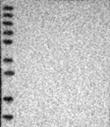
Analysis performed using a standard panel of samples. No bands detected.
Uncertain
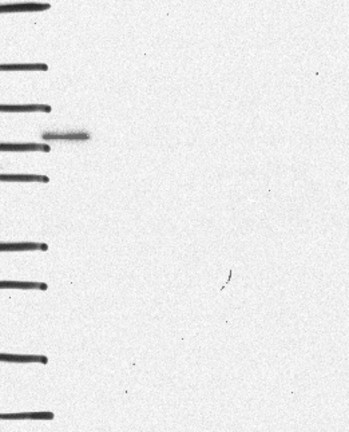
Analysis performed using a standard panel of samples. Single band differing more than +/-20% from predicted size in kDa and not supported by experimental and/or bioinformatic data.
Uncertain
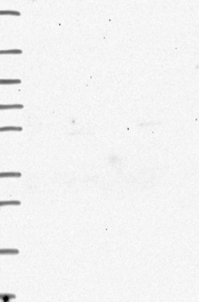
Analysis performed using a standard panel of samples. No bands detected.
Figure description
Lane 1: Marker [kDa] 230, 130, 95, 72, 56, 36, 28, 17, 11 Lane 2: RT4 Lane 3: U-251 MG Lane 4: Human Plasma Lane 5: Liver Lane 6: Tonsil
Lane 1: Marker [kDa] 250, 130, 95, 72, 55, 36, 28, 17, 10 Lane 2: RT4 Lane 3: U-251 MG Lane 4: Human Plasma Lane 5: Liver Lane 6: Tonsil
Lane 1: Marker [kDa] 250, 130, 95, 72, 55, 36, 28, 17, 10 Lane 2: RT4 Lane 3: U-251 MG Lane 4: Human Plasma Lane 5: Liver Lane 6: Tonsil
Target mass (kDa)
29
29, 21.8
29, 27.6, 21.8, 18.4, 15.4, 14.4, 11.6
Antibody dilution
1:250
1:270
1:170
PROTEIN ARRAY
Antibody HPA028930
Antibody HPA053429
Antibody HPA058961
Standard validation
Approved
Pass with quality comment low specificity (binding to 1-2 PrESTs >15% and <40%).
Supported
Pass with single peak corresponding to interaction only with its own antigen.
Supported
Pass with single peak corresponding to interaction only with its own antigen.
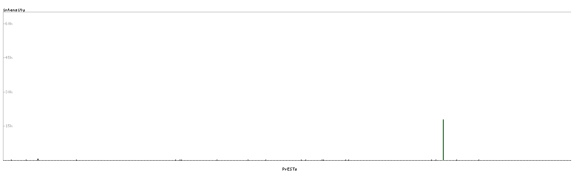
Figure description
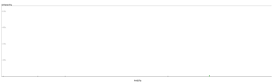
Antibody specificity analysis with protein arrays. Predicted and matching interactions are shown in green.
Antibody specificity analysis with protein arrays. Predicted and matching interactions are shown in green.
Antibody specificity analysis with protein arrays. Predicted and matching interactions are shown in green.
Antibody dilution
1:500
1:2150
1:2100
ANTIGEN INFORMATION
Antibody HPA028930
Antibody HPA053429
Antibody HPA058961
Antigen
Recombinant protein fragment
Recombinant protein fragment
Recombinant protein fragment
Length (aa)
102
49
35
Antigen sequence
SVGSVTSRPSTPTLGTPTPQTMSVSTKVGTPMSLTGQRFTVQMPTSQSPA
VKASIPATSAVQNVLINPSLIGSKNILITTNMMSSQNTANESSNALKRKR
ED