Heart muscle
Heart muscle
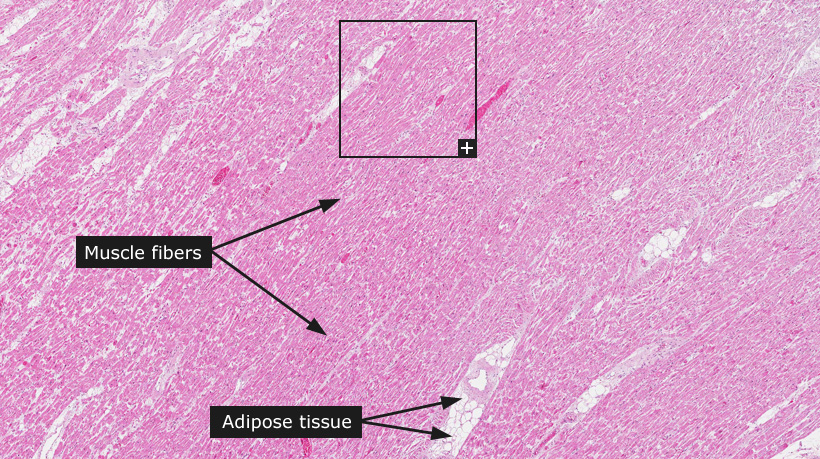
The human heart is in essence a coordinated muscle that flushes 6,000 liters of blood through the body daily during a lifetime. The underlying heart muscle is highly vascularized and under nervous control to set the pace of heart beats. The cardiac muscle (myocardium) together with the skeletal muscles are composed of striated muscle tissue that form parallel muscle fibers. Different to skeletal muscle, cardiac muscle cells may be branched instead of organized as parallel linear fibers. Striated muscle tissue of heart consists of cardiomyocytes arranged in fibers that exhibit cross-striations formed by alternating segments of thick and thin protein filaments.
The major cell type is cardiomyocytes, which usually contain one or two nuclei. The cells are rich in mitochondria and to a large extent contain actin and myosin proteins arranged in a repeating unit called a sarcomere. Histologically, this highly structured arrangement of sarcomeres appears as dark (A-bands) and light (I-bands) bands, which are clearly visible in the microscopic image. In addition to the muscle fibers, the myocardium also includes streaks of connective and cuffs of adipose tissue surrounding the smaller vessels. The cardiac muscle tissue is highly vascularized through the feeding coronary arteries that branch into smaller vessels that end in a dense network of capillaries running in between the fibers.
|