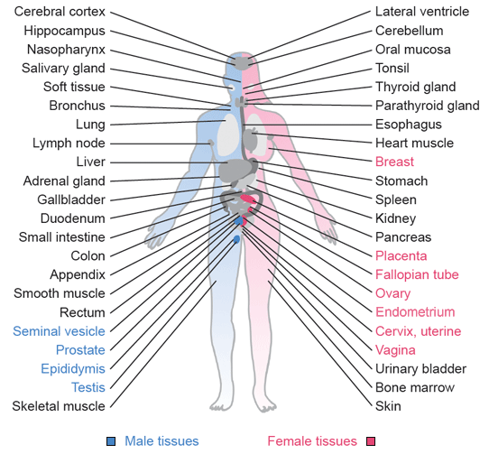
The normal histology section of the dictionary is based on representative sections from human tissues. Tissues have been collected from clinical specimens sent to the Department of Pathology at the Uppsala Akademiska hospital, Uppsala, Sweden for diagnostics. All specimens have been collected with consent of patients. All samples have been anonymized in accordance with ethical approval. Examples of normal tissue histology have been selected from regions in these surgical specimens where morphology appears as normal.
Tissues used in the dictionary have been formalin fixed, paraffin embedded and cut in 4 µm thin sections that have been placed on glass slides. Glass slides containing tissue sections have been stained using hematoxylin-eosin (HE). Hematoxylin stains cell nuclei blue and eosin stains the cytoplasm and membrane of cells together with connective tissue and extracellular matrix in various shades of red color. Following HE-staining, the sections of tissue have been scanned to obtain high-quality images corresponding to 40x magnification in a microscope.
Histology is defined as the study of microscopical anatomy of cells. Assessment of histology provides important and basic information for our understanding of biology and medicine. Histology is also an integral part of pathology and microscopy-based diagnostics. Histological structures, in terms of pathological consideration, are thus important to recognize and relate to when distinguishing a particular disease from normal. In particular inter-individual variations of the norm (for example related to age) can present a challenge to distinguish normal from a pathological condition.
|