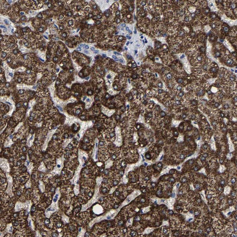
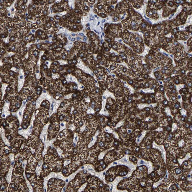
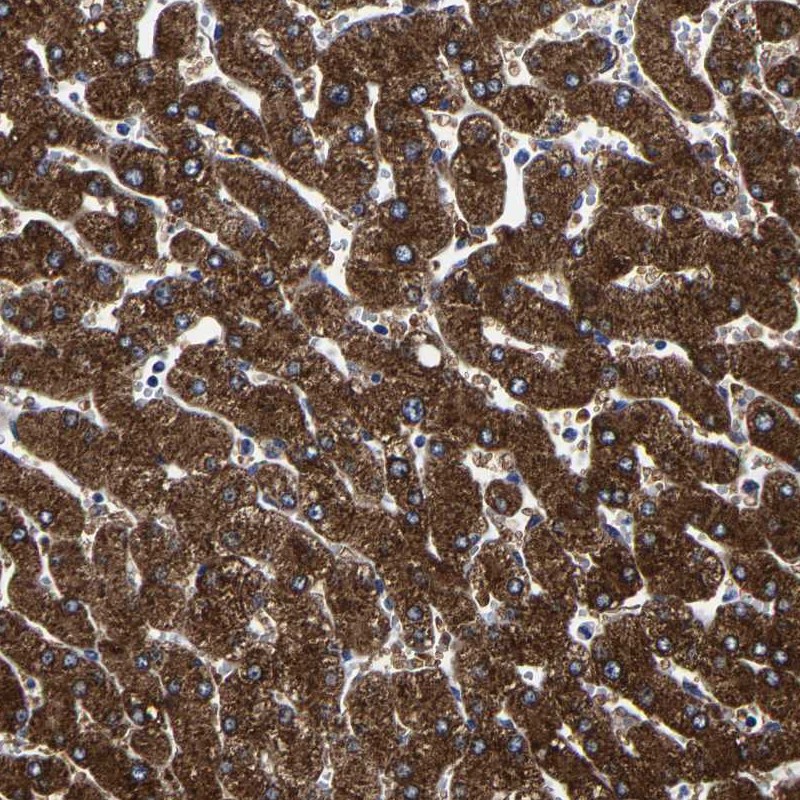
TISSUE
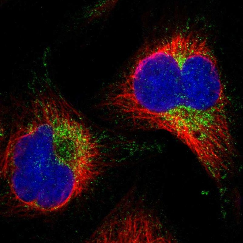
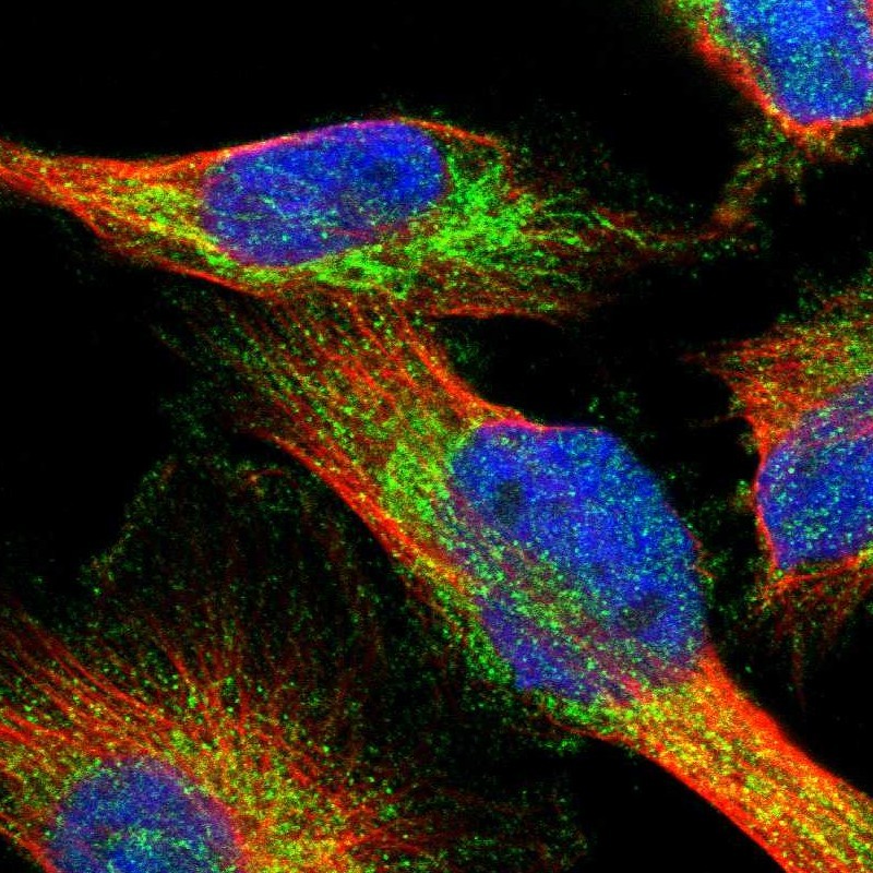
CELL
PATHOLOGY
ANTIBODY INFORMATION
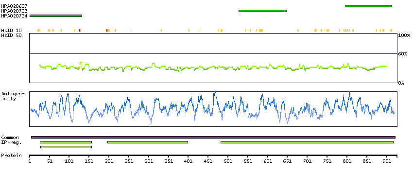
Antibody HPA020637
Antibody HPA020728
Antibody HPA020734
Provider
Product name
Host species
Clonality
Purity
Other gene match
Released in version
References
Proper citation
VALIDATION SUMMARY
IMMUNOCYTOCHEMISTRY
Formal validation: Independent
Standard validation
Figure description
Antibody dilution
Literature conformity
IMMUNOHISTOCHEMISTRY
Expression
Retrieval
RNA consistency
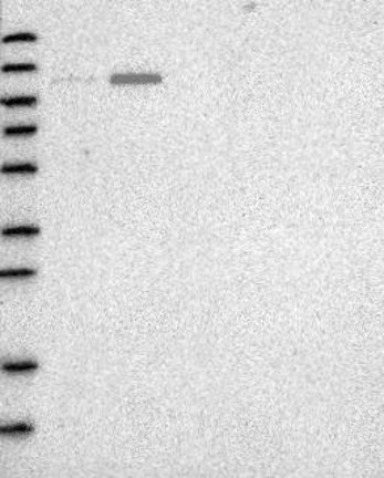
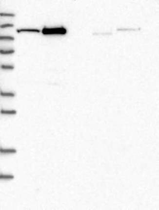
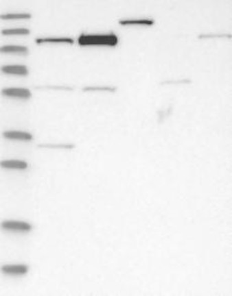
WESTERN BLOT
Target mass (kDa)
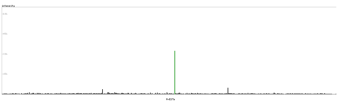
PROTEIN ARRAY
ANTIGEN INFORMATION
Antigen
Length (aa)
Antigen sequence
AEWLGLLGDEQVPQAESILDALSKHLVMKLSYGPEEKDMIVMRDSFGIRH
PSGHLEHKTIDLVAYGDINGFSAMAKTVGLPTAMAAKMLLDGEIGAKGLM
GPFSKEIYGPILERIKA
PVSMDICKQEEKLGFLVAKQDLVISLLPYVLHPLVAKACITNKVNMVTAS
YITPALKELEKSVEDAGITIIGELGLDPGLDHMLAMETIDKAKEVGATIE
SYISYCGGLPAPEHSNNPLRYK
MLQVHRTGLGRLGVSLSKGLHHKAVLAVRREDVNAWERRAPLAPKHIKGI
TNLGYKVLIQPSNRRAIHDKDYVKAGGILQEDISEACLILGVKRPPEEKL
MSRKTYAFFSHTIKAQEANMGLLDEILKQEIR
Matching transcripts
RELEVANT PUBLICATIONS
1.