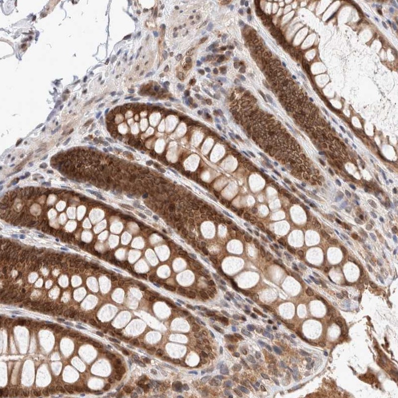
TISSUE
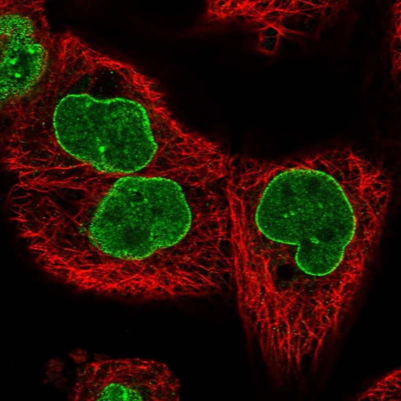
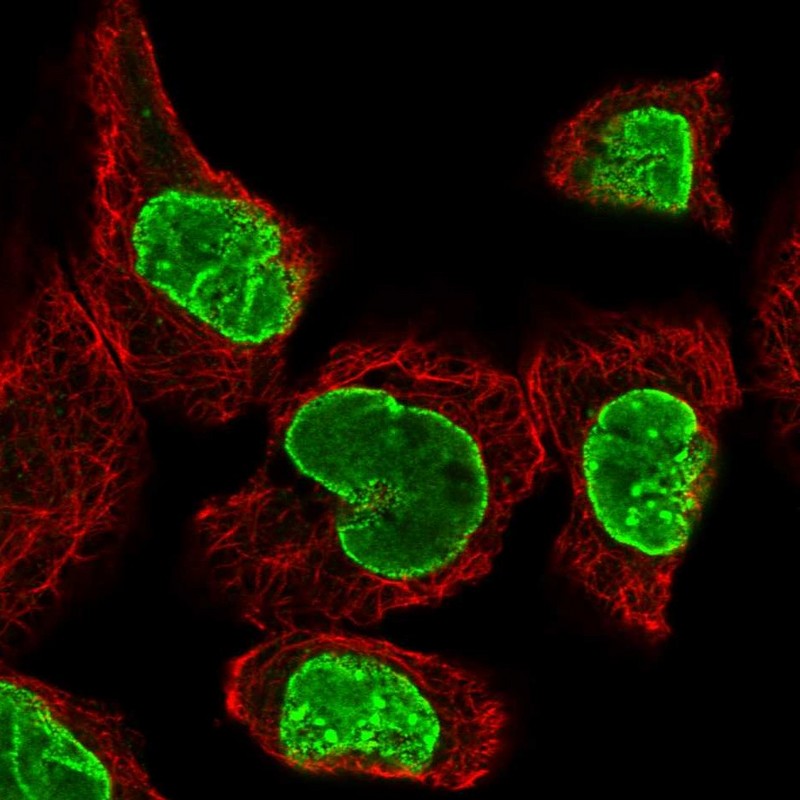
CELL
PATHOLOGY
ANTIBODY INFORMATION
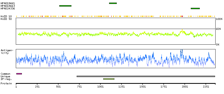
Antibody HPA019661
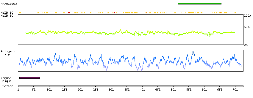
Antibody HPA019663
Antibody HPA024336
Provider
Product name
Host species
Clonality
Purity
Other gene match
Released in version
References
Proper citation
VALIDATION SUMMARY
IMMUNOCYTOCHEMISTRY
Formal validation: Independent
Standard validation
Figure description
Antibody dilution
Literature conformity
IMMUNOHISTOCHEMISTRY
Expression
Retrieval
RNA consistency
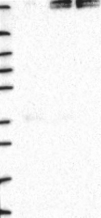
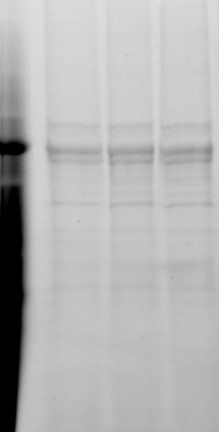
WESTERN BLOT
Formal validation: Genetic
Target mass (kDa)
Loading control
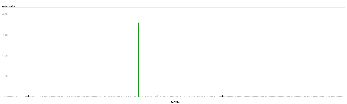
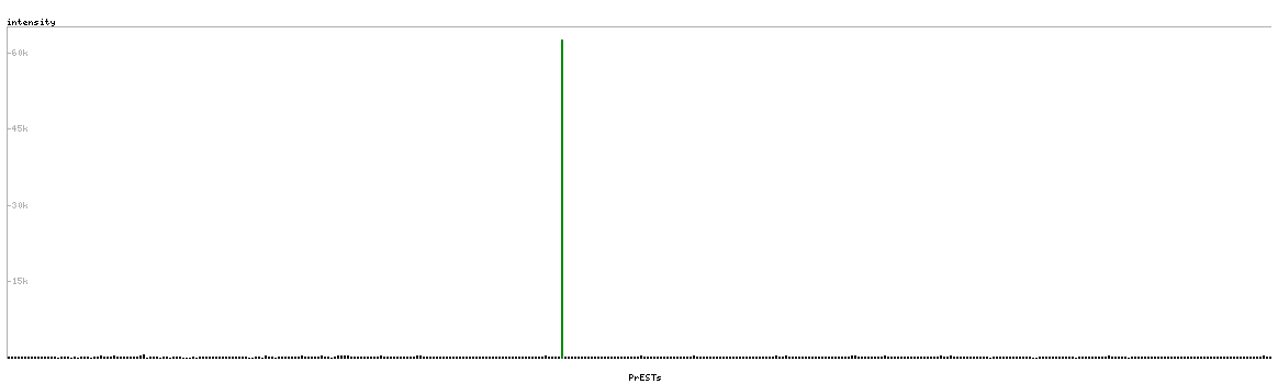
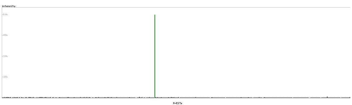
PROTEIN ARRAY
ANTIGEN INFORMATION
Antigen
Length (aa)
Antigen sequence
KMASVRQHLEETTQKAESQLLECKASWEERERMLKDEVSKCVCRCEDLEK
QNRLLHDQIEKLSDKVVASVKEGVQGPLNVSLSEEGKSQE
RDEEVSSADISSSSEVISQHLVSYRNIEELQQQNQRLLVALRELGETRER
EEQETTSSKITELQLKLESALTELEQLRKSRQHQMQLVDSIVRQRDMYRI
LLSQTTGVAIPLHASSLDDVSLASTPKRPSTSQTVSTPAP
RRPPHPLPPRLTIHAPPQELGPPVQRIQMTRRQSVGRGLQLTPGIGGMQQ
HFFDDEDRTVPSTPTLVVPHRTDGFAEAIHSPQVAGVPRFRFGPPEDMPQ
TSSSHS
Matching transcripts


