We use cookies to enhance the usability of our website. If you continue, we'll assume that you are happy to receive all cookies. More information. Don't show this again.
Antibody staining overlaps with GFP tagged protein
Validated
Antibody staining overlaps with GFP tagged protein
Figure description
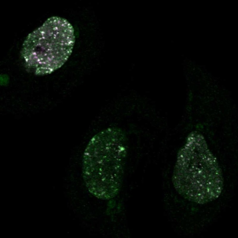
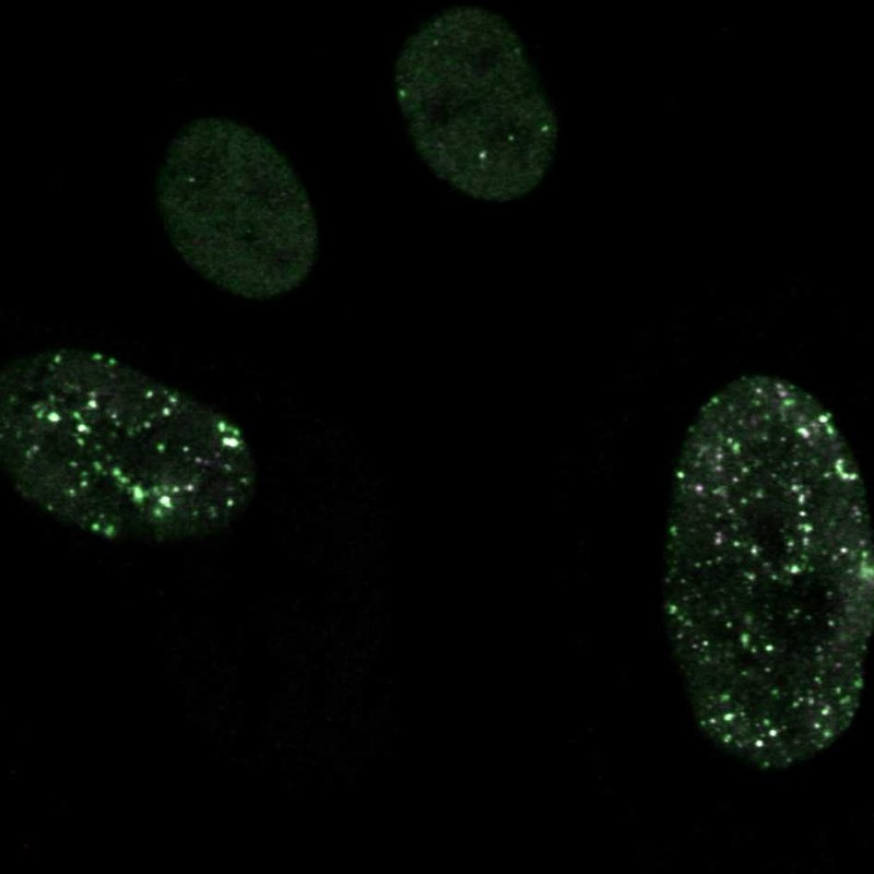
Immunofluorescent staining of transgenic HeLa cells show antibody staining in nuclear bodies & nucleoplasm and GFP expression in nuclear bodies & nucleoplasm.
Immunofluorescent staining of transgenic HeLa cells show antibody staining in nuclear bodies & nucleoplasm and GFP expression in nuclear bodies & nucleoplasm.
Antibody dilution
1:29
1:37
Formal validation: Independent
Validated
Antibody staining overlaps with antibody HPA008752.
Validated
Antibody staining overlaps with antibody HPA006724.
Standard validation
Supported
Supported
Figure description
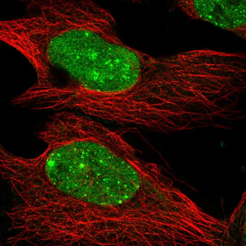
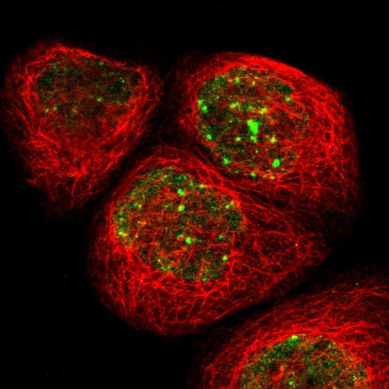
Immunofluorescent staining of human cell line U-2 OS shows localization to nucleoplasm & nuclear bodies.
Immunofluorescent staining of human cell line A-431 shows localization to nucleoplasm & nuclear bodies.
Antibody dilution
1:29
1:37
Literature conformity
The subcellular location is supported by literature.
The subcellular location is supported by literature.
IMMUNOHISTOCHEMISTRY
Antibody HPA006716
Antibody HPA006724
Antibody HPA008752
Standard validation
Supported
Supported
Figure description
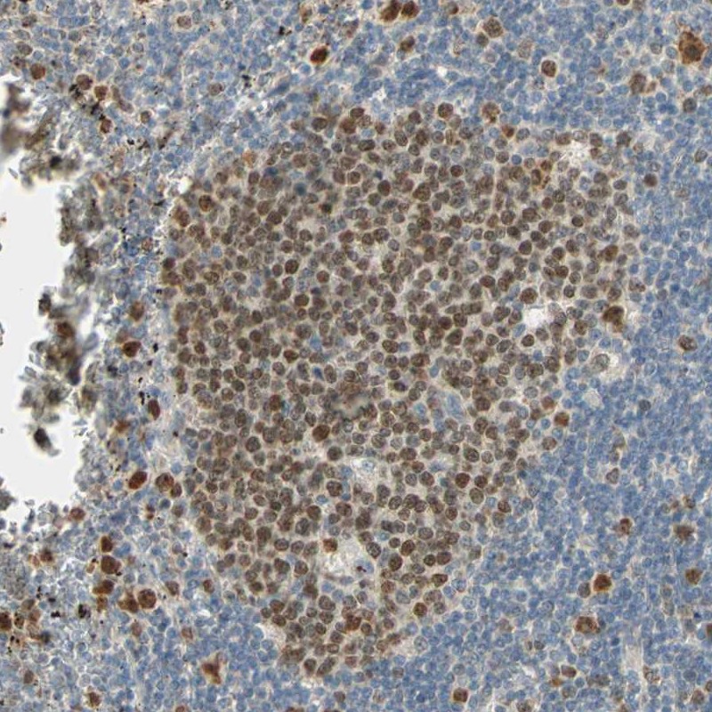
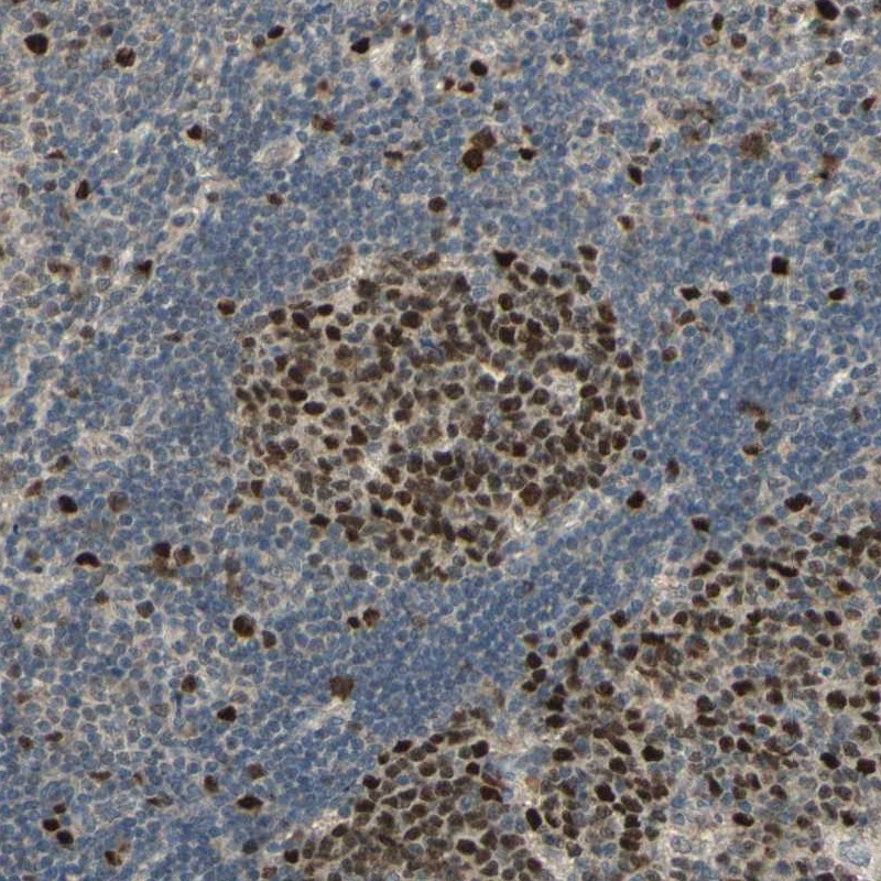
Immunohistochemical staining of human lymph node shows distinct nuclear positivity in germinal center cells.
Immunohistochemical staining of human lymph node shows distinct nuclear positivity in germinal center cells.
Expression
RNA: detected in 35 tissues Protein: detected in 44 cell types
RNA: detected in 35 tissues Protein: detected in 58 cell types
Retrieval
HIER pH6
HIER pH6
Antibody dilution
1:200
1:10
Literature conformity
Partly consistent with extensive gene/protein characterization data.
Consistent with extensive gene/protein characterization data.
RNA consistency
Mainly consistent with RNA expression data.
Mainly consistent with RNA expression data.
Standard validation
Approved
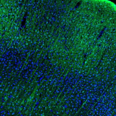
Figure description
Predominant (although not exclusive) distribution in neuronal somata and processes from various layers of the cerebral cortex.
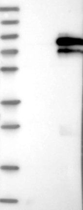
WESTERN BLOT
Antibody HPA006716
Antibody HPA006724
Antibody HPA008752
Standard validation
Supported
Analysis performed using a standard panel of samples. Band of predicted size in kDa (+/-20%) with additional bands present.
Uncertain
Analysis performed using a standard panel of samples. Weak band of predicted size but with additional bands of higher intensity also present.
Supported
Band of predicted size in kDa (+/-20%) with additional bands present.
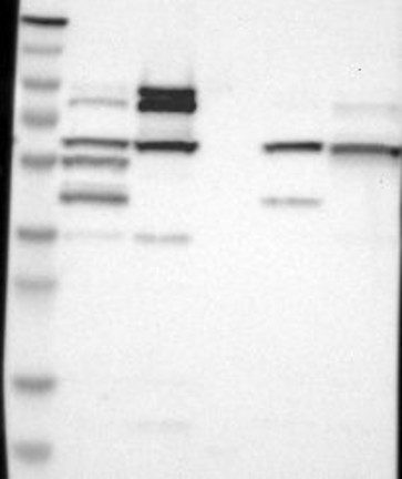
Figure description
Lane 1: Marker [kDa] 230, 130, 95, 72, 56, 36, 28, 17, 11 Lane 2: RT4 Lane 3: U-251 MG Lane 4: Human Plasma Lane 5: Liver Lane 6: Tonsil
Lane 1: Marker [kDa] 250, 130, 95, 72, 55, 36, 28, 17, 10 Lane 2: Negative control (vector only transfected HEK293T lysate) Lane 3: Over-expression Lysate (Co-expressed with a C-terminal myc-DDK tag (~3.1 kDa) in mammalian HEK293T cells, LY402768)
Target mass (kDa)
56.2
56.2
56.2
Antibody dilution
1:250
1:250
1:250
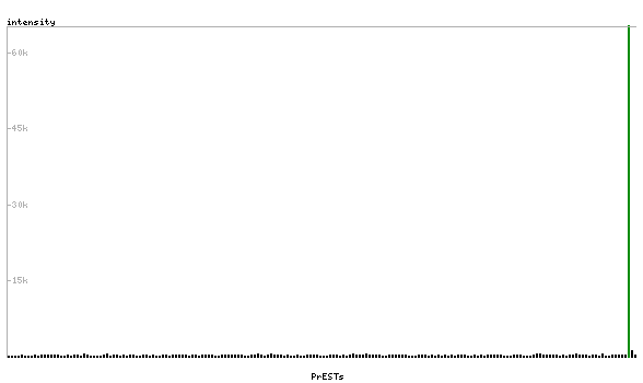
PROTEIN ARRAY
Antibody HPA006716
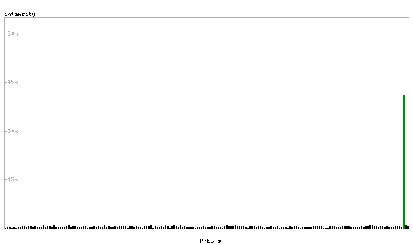
Antibody HPA006724
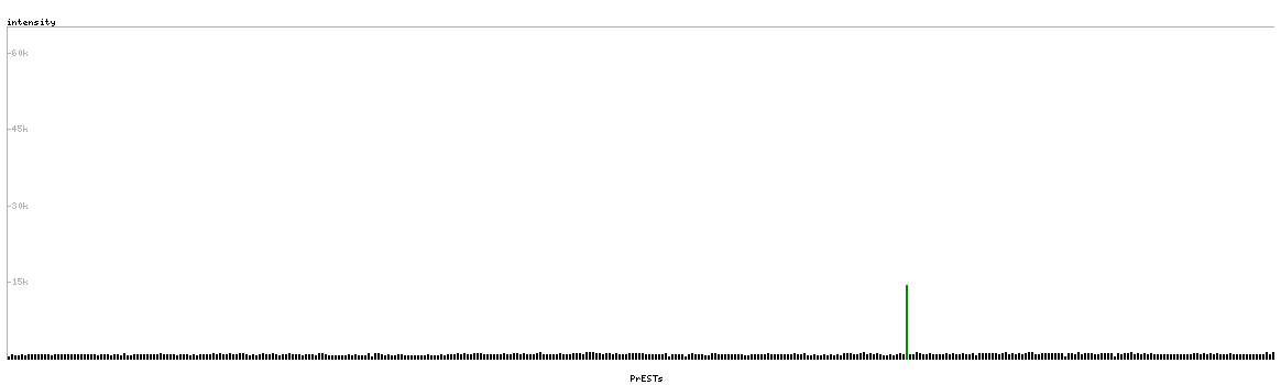
Antibody HPA008752
Standard validation
Supported
Pass with single peak corresponding to interaction only with its own antigen.
Supported
Pass with single peak corresponding to interaction only with its own antigen.
Supported
Pass with single peak corresponding to interaction only with its own antigen.
Figure description
Antibody specificity analysis with protein arrays. Predicted and matching interactions are shown in green.
Antibody specificity analysis with protein arrays. Predicted and matching interactions are shown in green.
Antibody specificity analysis with protein arrays. Predicted and matching interactions are shown in green.