TISSUE
CELL
PATHOLOGY
ANTIBODY INFORMATION
Antibody HPA040025
Antibody HPA043926
Antibody HPA061524
Antibody CAB002433
Provider
Product name
Host species
Clonality
Purity
Other gene match
Released in version
References
Proper citation
VALIDATION SUMMARY
IMMUNOCYTOCHEMISTRY
Formal validation: Independent
Standard validation
Figure description
Antibody dilution
Literature conformity
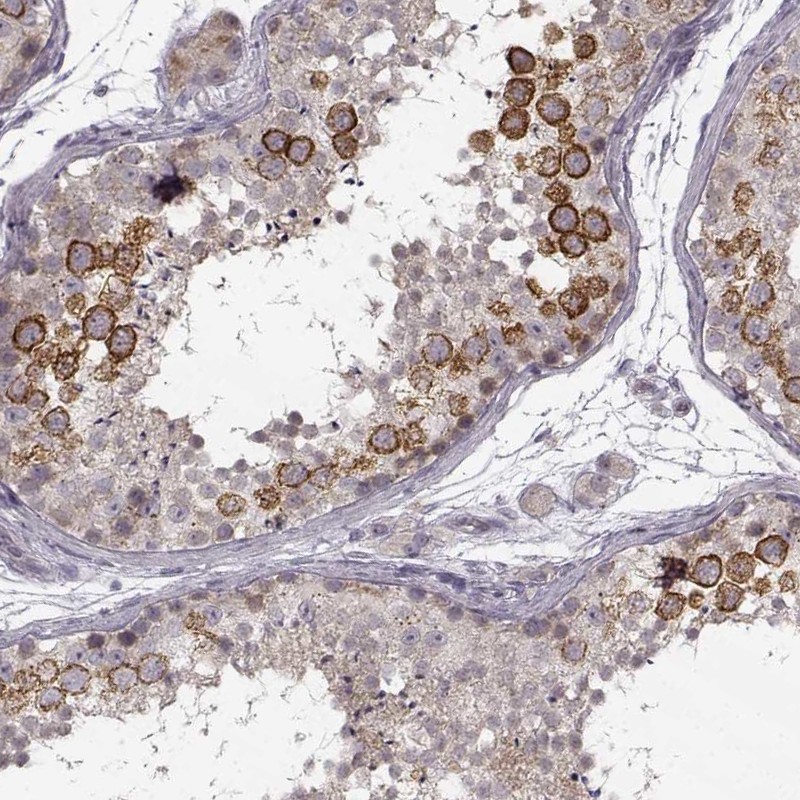
IMMUNOHISTOCHEMISTRY
Formal validation: Orthogonal
Expression
Retrieval
RNA consistency
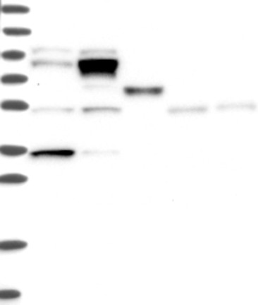
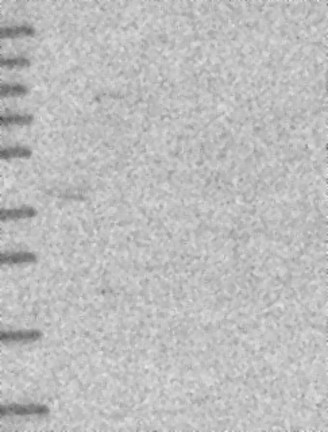
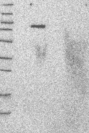
WESTERN BLOT
Target mass (kDa)
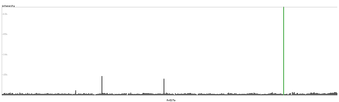
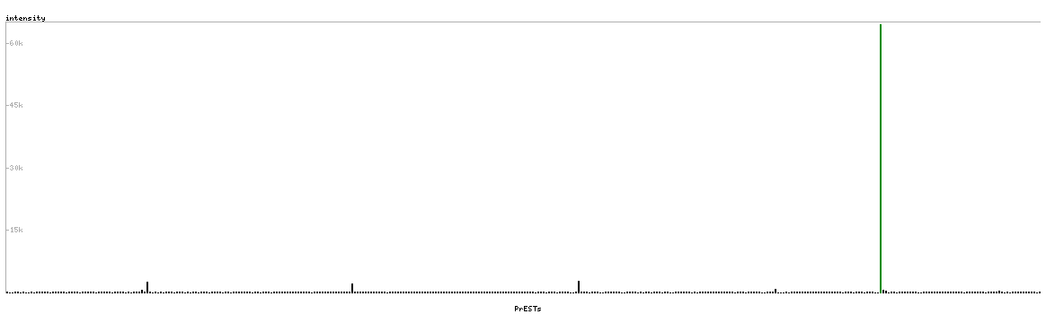
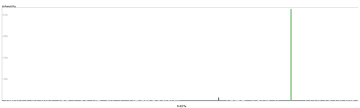
PROTEIN ARRAY
ANTIGEN INFORMATION
Antigen
Length (aa)
Antigen sequence
SSAAHTQATLLLQEKYDSMVQSLEDVTAQFESYKALTASEIEDLKLENSS
LQEKVAKAGKNAEDVQHQILATESSNQEYVRMLLDLQTKSALKET
KRFNDPSGCAPSPGAYDVKTLEVLKGPVSFQKSQRFKQQKESKQNLNVDK
DTTLPASARKVKSSESKKESQKNDKDLKILEKEIRVLLQERGAQ
LKQQEEDFRKQLEDEEGRKAEKENTTAELTEEINKWRLLYEELYNKTKPF
QLQLDAFEVEKQALLNEHGAAQEQLNKIRDSYAKLLGHQ
Matching transcripts