TISSUE
CELL
PATHOLOGY
ANTIBODY INFORMATION
Antibody HPA004817
Antibody HPA061417
Antibody CAB013460
Provider
Product name
Host species
Clonality
Purity
Other gene match
Released in version
References
Proper citation
VALIDATION SUMMARY
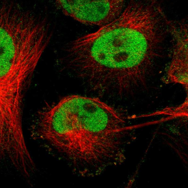
IMMUNOCYTOCHEMISTRY
Standard validation
Figure description
Antibody dilution
Literature conformity
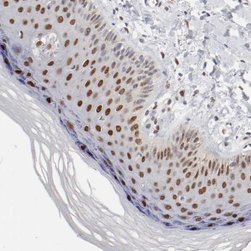
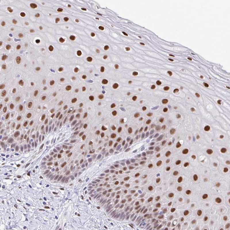
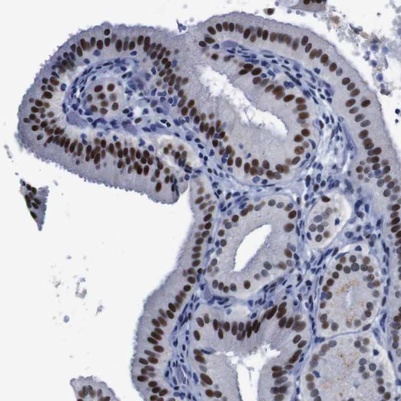
IMMUNOHISTOCHEMISTRY
Formal validation: Orthogonal
Formal validation: Independent
Expression
Retrieval
RNA consistency
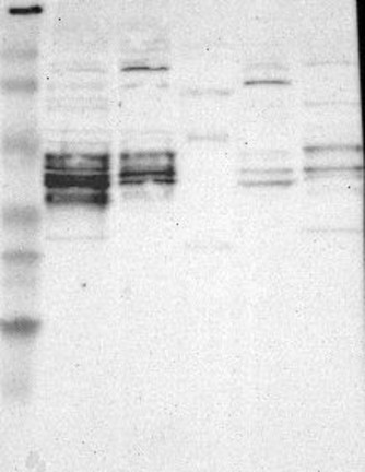
WESTERN BLOT
Target mass (kDa)
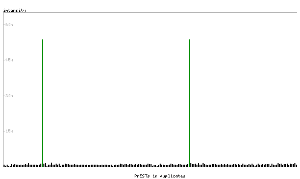
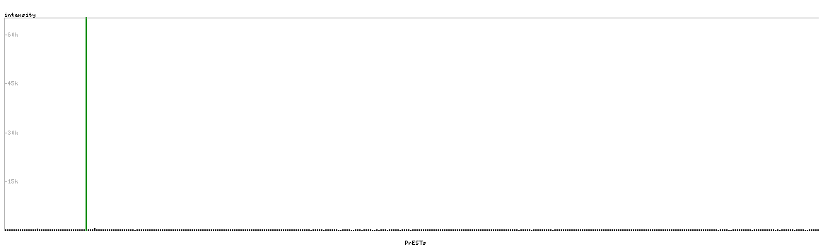
PROTEIN ARRAY
ANTIGEN INFORMATION
Antigen
Length (aa)
Antigen sequence
EKLEFMLVAHGPVCKISPEERRSPPAPGLQPMRSGGGSVGAVVVKQEPLE
EDSPSSSSAGLDKAQRSVIKPISIAGGFYGEEPLHTPIVVTSTPAVTPGT
SNLVFTYPSVLEQESPASPSESCSKAHRRSSSSGDQSSDSL
WMVQPTVITSMSNPYPRSHPYSPLPGLASVPGHMALPRPGVIKTIGTTVG
RRRRDEQLS
Matching transcripts