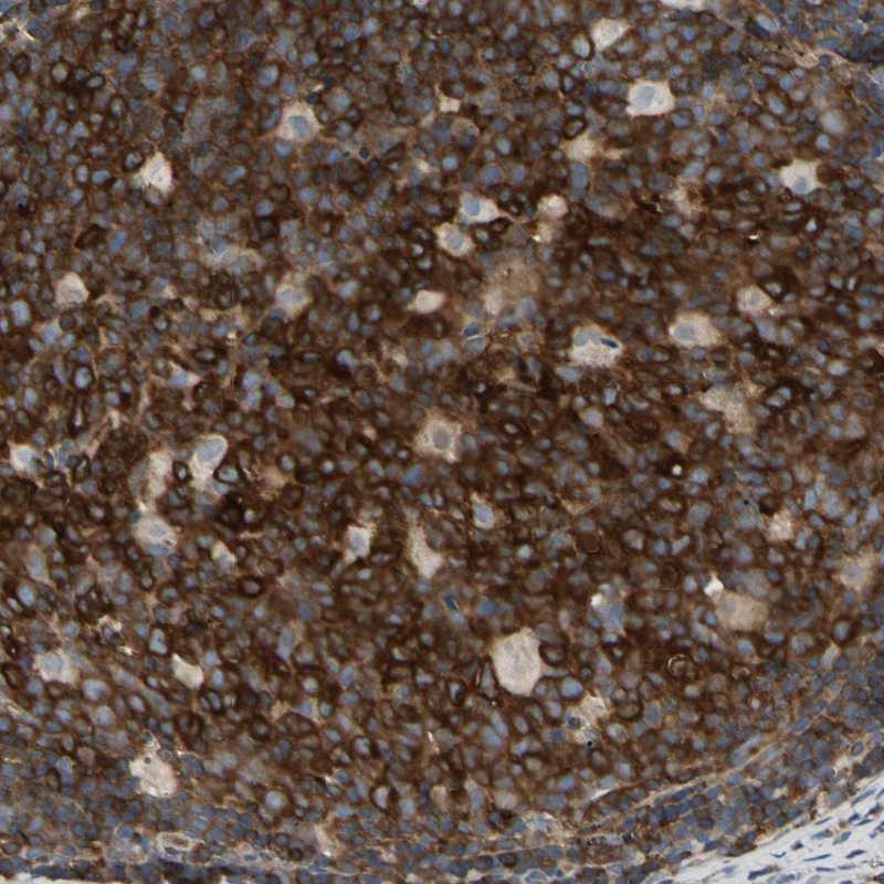
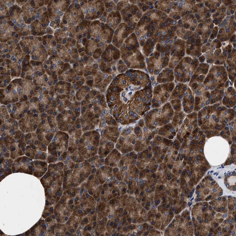
TISSUE
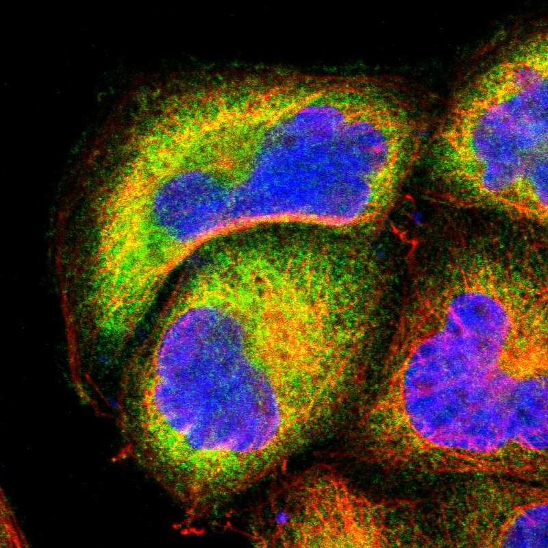
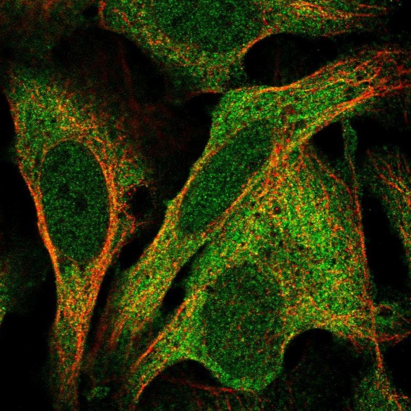
CELL
PATHOLOGY
ANTIBODY INFORMATION
Antibody HPA008422
Antibody HPA023900
Antibody CAB022098
Provider
Product name
Host species
Clonality
Purity
Other gene match
Released in version
References
Proper citation
VALIDATION SUMMARY
IMMUNOCYTOCHEMISTRY
Standard validation
Figure description
Antibody dilution
Literature conformity
IMMUNOHISTOCHEMISTRY
Formal validation: Orthogonal
Formal validation: Independent
Expression
Retrieval
RNA consistency
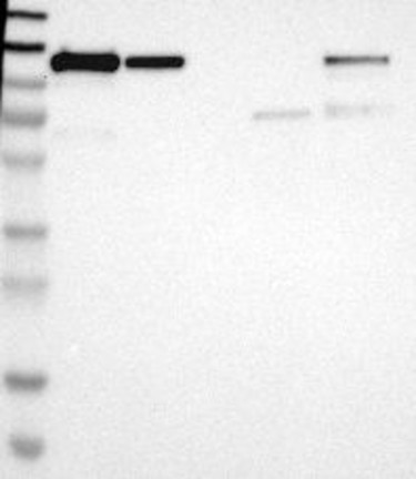
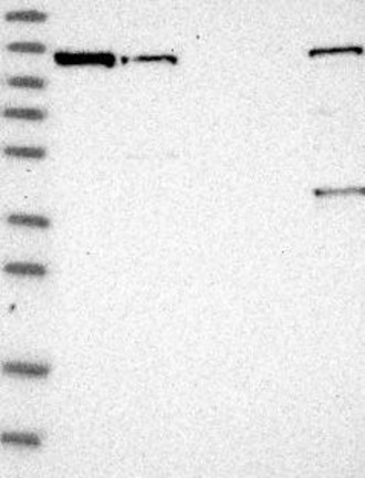
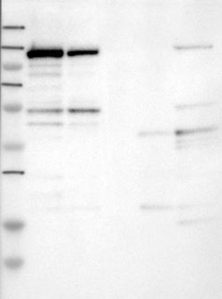
WESTERN BLOT
Target mass (kDa)
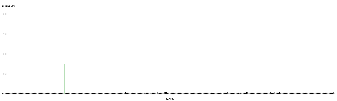
PROTEIN ARRAY
ANTIGEN INFORMATION
Antigen
Length (aa)
Antigen sequence
LLNAAQNTMEPPLTPPSPAGPGLSLGDTALQNLEQLLDGPEAQGSWAELA
ERLGLRSLVDTYRQTTSPSGSLLRSYELAGGDLAGLLEALSDMGLEEGVR
LLRGPETRDKLPSTEVKEDSAYGSQSVEQEA
ICNYEGPAKIEVDLVTHSDPPRAHAHSLVGKQCSELGICAVSVGPKDMTA
QFNNLGVLHVTKKNMMGTMIQKLQRQRLRSRPQGLTEAEQRELEQEAKEL
KKVMDLSIVRLRFSAFLRASDGSFSLPLKPVISQP
Matching transcripts
RELEVANT PUBLICATIONS
1.