TISSUE
CELL
PATHOLOGY
ANTIBODY INFORMATION
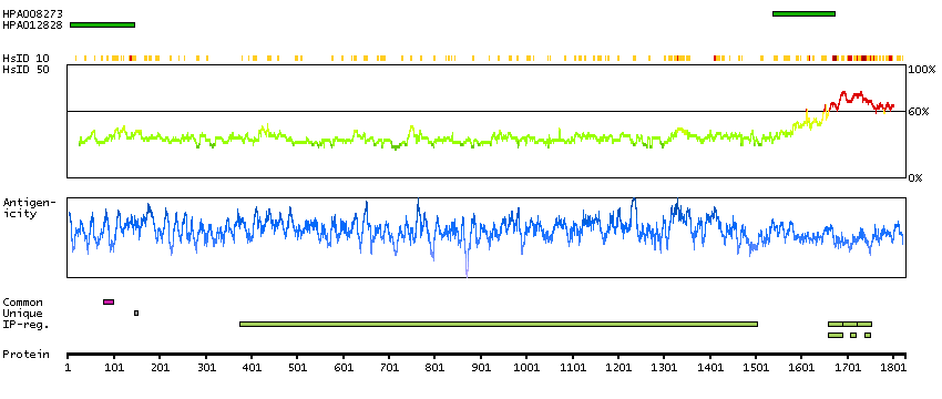
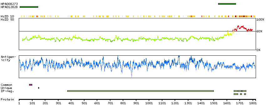
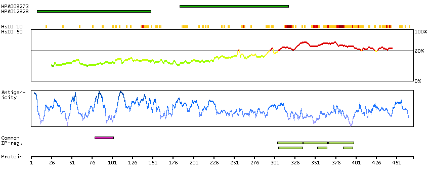
Antibody HPA008273
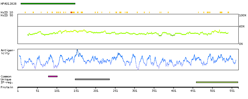
Antibody HPA012828
Antibody CAB001984
Provider
Product name
Host species
Clonality
Purity
Other gene match
Released in version
References
Proper citation
VALIDATION SUMMARY
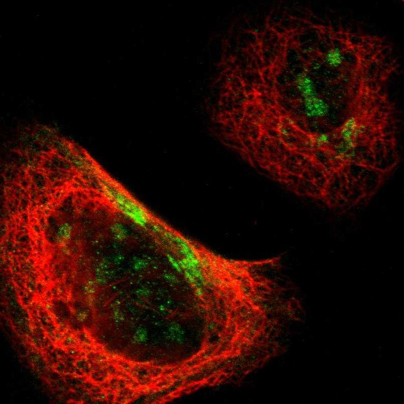
IMMUNOCYTOCHEMISTRY
Formal validation: Independent
Standard validation
Figure description
Antibody dilution
Literature conformity
IMMUNOHISTOCHEMISTRY
Formal validation: Orthogonal
Expression
Retrieval
RNA consistency
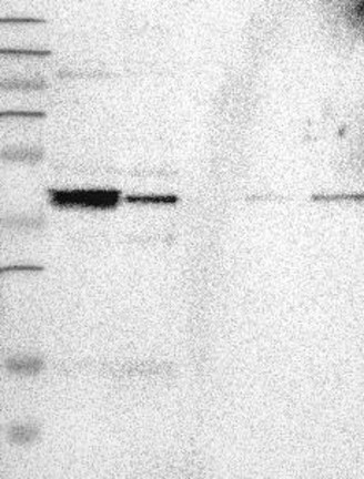
WESTERN BLOT
Target mass (kDa)
PROTEIN ARRAY
ANTIGEN INFORMATION
Antigen
Length (aa)
Antigen sequence
SLPRPSSILPPRRGVSGDRDENSFSLNSSISSSARRTTRSEPIRRAGKSG
TSTPTTPGSTAITPGTPPSYSSRTPGTPGTPSYPRTPHTPGTPKSAILVP
SEKKVAIIRTPPKSPATPKQLRLINQPLPDLKNVK
EAKAPHWTSAPLTEASAHSHPPEIKDQGGAGEGLVRSANGFPYREDEEGA
FGEHGSQGTYSNTKENGINGELTSADRETAEEVSARIVQVVTAEAVAVLK
GEQEKEAQHKDQTAALPLAAEETANLPPSPPPSPASEQTVT
Matching transcripts
RELEVANT PUBLICATIONS
1.
2.