We use cookies to enhance the usability of our website. If you continue, we'll assume that you are happy to receive all cookies. More information. Don't show this again.
Fields »
 

Field

Term
Gene name

Class

Subclass

Chromosome

External id

In atlas

Reliability

Reliability

Reliability

Validation

Method

Validation

Method

Validation

Method

Validation

Location

Searches

Tissue

Cell type

Expression

Tissue

Specificity

Cell line

Specificity

Cancer

Specificity

Tissue

Detectable

Cell line

Detectable

Cancer

Detectable

Cancer

Prognosis

Category

Score

Score

Score

Antibodies

Column


ABOUT
HELP
BLOG
  • THE HUMAN PROTEOME
  • THE HUMAN TISSUES
  • THE HUMAN CELL
  • HUMAN PATHOLOGY
  • PROTEIN CLASSES
  • PROTEIN EVIDENCE
    • NEWS
    • BLOG
    • EVENTS
    • PRESS ROOM
    • LEARN
    • DICTIONARY
    • METHODS
    • CELL LINES
  • THE PROJECT
  • INTRODUCTION
  • ORGANIZATION
  • PUBLICATIONS
  • PUBLICATION DATA
  • ANTIBODY SUBMISSION
  • ANTIBODY AVAILABILITY
  • LINKS
  • CONTACT
  • TECHNICAL DATA
  • ANTIBODY VALIDATION
  • ASSAYS & ANNOTATION
  • DISCLAIMER
  • DOWNLOADABLE DATA
  • HELP & FAQ
  • LICENCE & CITATION
  • PRIVACY STATEMENT
  • RELEASE HISTORY
CD79A
  • premium

    TISSUE

  • CELL

  • PATHOLOGY

ANTIBODY INFORMATION VALIDATION SUMMARY IMMUNOCYTOCHEMISTRY IMMUNOHISTOCHEMISTRY WESTERN BLOT PROTEIN ARRAY ANTIGEN INFORMATION RELEVANT PUBLICATIONS

ANTIBODY INFORMATION


Antibody CAB000019


Provider

DakoCytomation
 

Product name

M7050  

Host species

Mouse  

Clonality

mAb  

Purity

Supernatant  

Other gene match


Released in version

1  

References


Proper citation

Dako Cat#M7050, RRID:AB_2244527  

VALIDATION SUMMARY


Antibody CAB000019

 
Immunohistochemistry


Validated
 
Western blot

Uncertain  

IMMUNOHISTOCHEMISTRY


Antibody CAB000019


Formal validation: Orthogonal

Validated

Pearson correlation >0.6 for protein and RNA expression in cell lines.


Figure description

Distribution of protein expression (antibody staining) and RNA expression (TPM) across 44 cell lines.  
 

Standard validation

Supported  


Figure description

Immunohistochemical staining of human lymph node shows strong cytoplasmic positivity in reaction center cells.  

Expression

RNA: detected in 31 tissues
Protein: detected in 12 cell types
 

Retrieval

HIER pH9  

Antibody dilution

1:500  

Literature conformity

Consistent with extensive gene/protein characterization data.  

RNA consistency

Consistent with RNA expression data.  
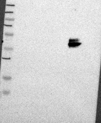
WESTERN BLOT


Antibody CAB000019


Standard validation

Uncertain

Analysis performed using a standard panel of samples.
Single band larger than predicted size in kDa (+20%) but partly supported by experimental and/or bioinformatic data.


Figure description

Lane 1: Marker [kDa] 250, 130, 95, 72, 55, 36, 28, 17, 11
Lane 2: RT4
Lane 3: U-251 MG
Lane 4: Human Plasma
Lane 5: Liver
Lane 6: Tonsil
 

Target mass (kDa)

27.9, 25, 20.8  

Antibody dilution

1:500  

ANTIGEN INFORMATION


Antibody CAB000019


Antigen

Recombinant protein  

Length (aa)


Antigen sequence


Matching transcripts


Other gene match


Contact

  • BLOG
  • PRESS ROOM
  • contact@proteinatlas.org

The Project

  • INTRODUCTION
  • ORGANIZATION
  • PUBLICATIONS
  • DOWNLOADABLE DATA

The Human Protein Atlas

  • ANTIBODY AVAILABILITY
  • LICENCE & CITATION
  • HELP & FAQ
The Human Protein Atlas project is funded
by the Knut & Alice Wallenberg foundation.