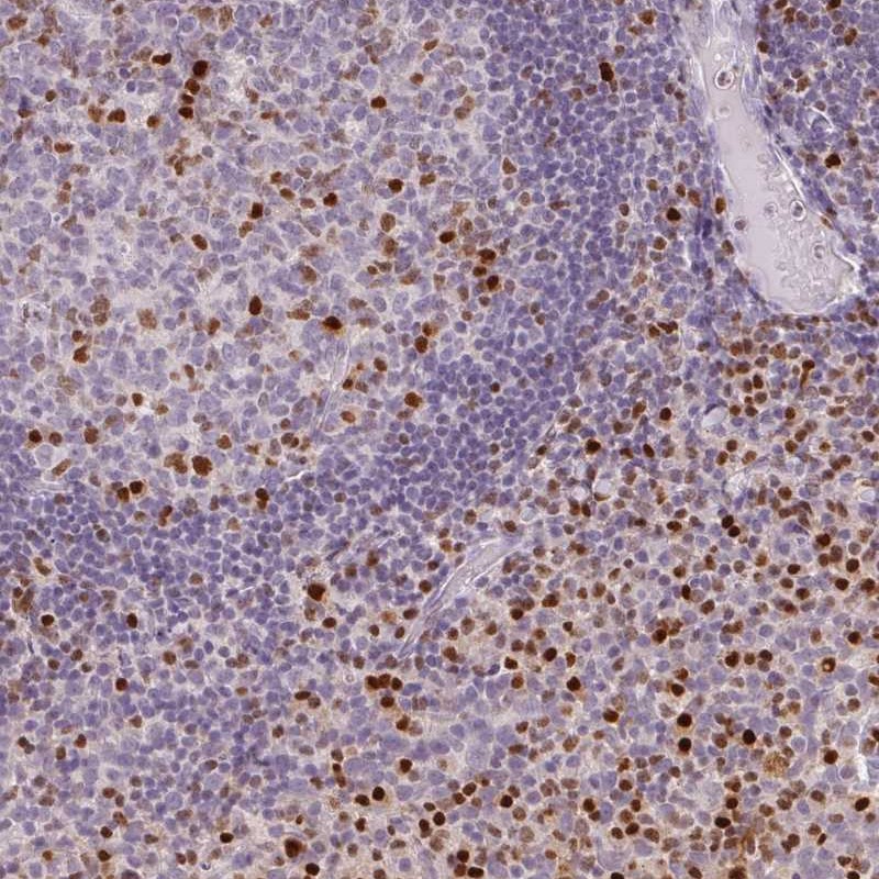
TISSUE
CELL
PATHOLOGY
ANTIBODY INFORMATION
Antibody HPA022040
Antibody CAB008374
Antibody CAB062561
Antibody CAB068226
Provider
Product name
Host species
Clonality
Purity
Other gene match
Released in version
References
Proper citation
VALIDATION SUMMARY
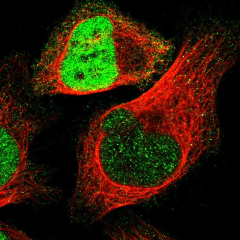
IMMUNOCYTOCHEMISTRY
Standard validation
Figure description
Antibody dilution
Literature conformity
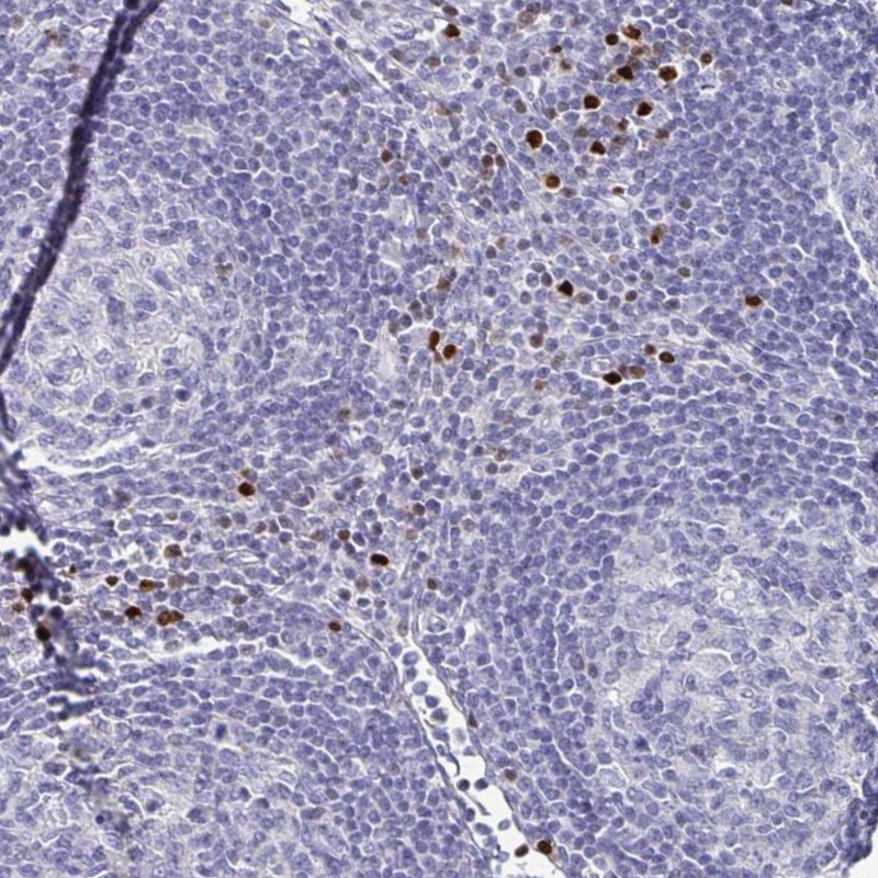
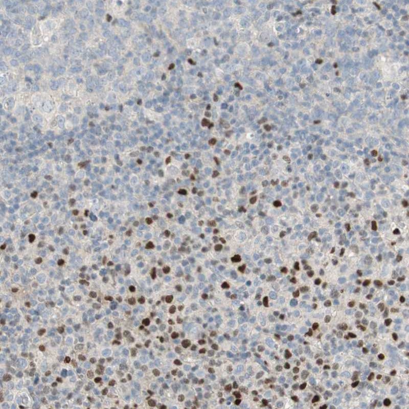
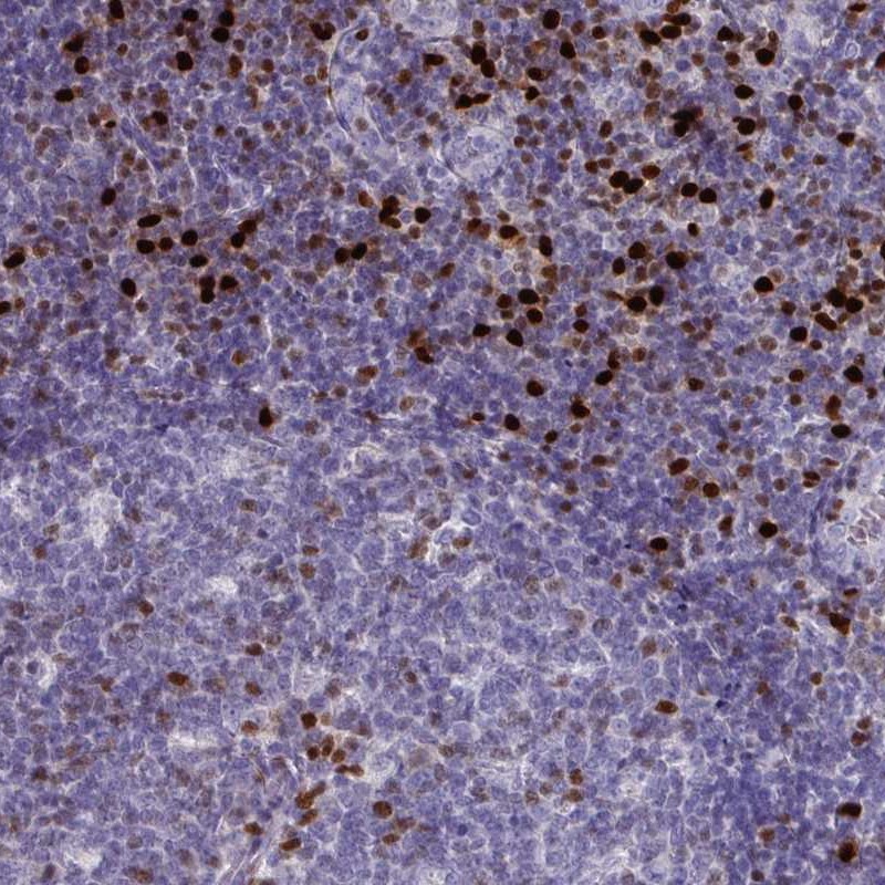
IMMUNOHISTOCHEMISTRY
Formal validation: Orthogonal
Expression
Retrieval
RNA consistency
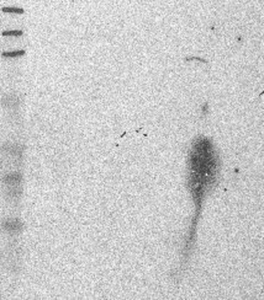
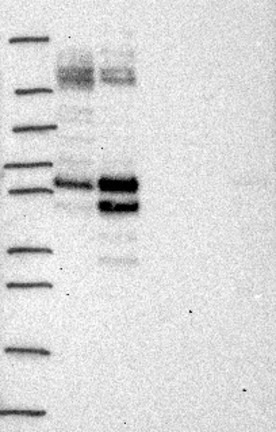
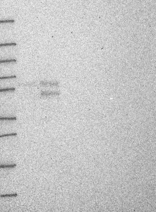
WESTERN BLOT
Target mass (kDa)
PROTEIN ARRAY
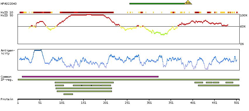
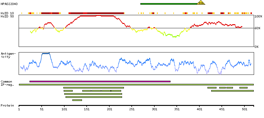
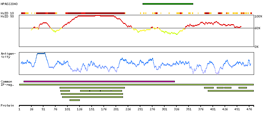
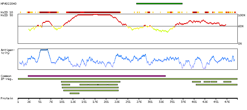
ANTIGEN INFORMATION
Antigen
Length (aa)
Antigen sequence
LNSAPSPFNPQGQSQITDPRQAQSSPPWSYDQSYPSYLSQMTSPSIHSTT
PLSSTRGTGLPAITDVPRRISGASELGPFSDPRQFPSISSLTESRFSNPR
MHYPA
Matching transcripts
RELEVANT PUBLICATIONS
1.
2.
3.
4.