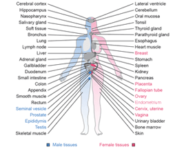
Oral mucosa
Oral mucosa
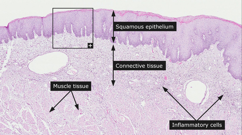
The surfaces in the oral cavity are lined with stratified squamous nonkeratinized epithelium. The multi-layered epithelium has protective function and protects the underlying structures from mechanical, chemical and thermal damages. Starting from the basement membrane the layers include a basal layer, a prickle cell layer and a stratum superficiale. The basal layer contains columnar cells that show mitotic activity and are responsible for cell renewal. Overlying the basal layer is the prickle cell layer (stratum spinosum) containing polygonal cells held together by desmosomes. The uppermost layer is the stratum superficiale that consists of flat squamous cells. The oral mucosa has a rich blood supply and in the underlying lamina propria mucosae there's loose connective tissue with numerous capillaries. Spread lymphocytes are also normally present in the lamina propria. Fingerlike protrusions of connective tissue, termed papillae, fold into the epithelial layer. Capillaries are lined by a single layer of endothelial cells, and have a surrounding basal lamina. Underlying the lamina propria is the submucosa with large collagen fibers and elastic fibers, attaching the mucosa to the muscle tissue beneath.
Cancer: Head and neck cancer
|