Lung cancer
Lung cancer is the leading cause of cancer-related death world-wide. Smoking is accepted as the major risk factor, responsible for 70-90% of all cancer cases, although the etiology of lung cancer appears multifactorial with both environmental and genetic factors playing a role. One important reason for the poor outcome of patients with lung cancer is that the disease is commonly diagnosed in advanced stages and therefore often not resectable by surgery. Symptoms of lung cancer are unspecific and dependent on anatomical site and include persistent coughing, hemoptysis, repeated pneumonia and signs of respiratory distress.
Lung cancer can be principally divided into small cell carcinoma and non-small cell carcinoma. Small cell lung carcinoma originates from neuroendocrine cells and accounts for approximately 15% of all primary lung cancers. This extremely rapidly proliferating cancer is generally treated with chemotherapy with initial good tumor response. However, after initial response, small cell lung carcinoma nearly always progresses, with only anecdotal long term survivors.
Non-small cell carcinoma is suggested to originate from bronchogenic or alveolar cells and represents the most common form of primary lung cancer. Patient therapy is based on the tumour extent. In principal, limited stage tumors are treated surgically, sometimes with the addition of chemotherapy and radiotherapy. Advanced stages are palliatively treated with a combination of cytotoxic drugs and recently developed targeted drugs. Unfortunately, often the effect of treatment leads to only modest survival prolongation.
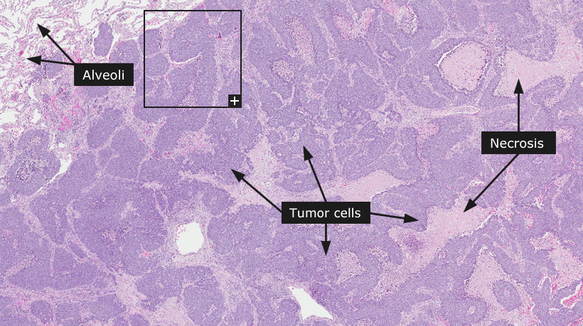
The histology-based classification has only minor impact on therapy and is under revision due to the identification of clinically relevant molecular aberrations and their relation to histopathological subtypes. Non-small cell carcinomas are classified as adenocarcinoma, squamous cell carcinoma or large cell carcinoma and very rare entities like pleomorphic carcinoma and mixed tumors. Necrosis, hemorrhage and abundant mitotic figures are common features in non-small cell lung carcinomas. As in several other forms of cancer, the infiltration of various inflammatory cells can be prominent and occaisional hisiocytic multinucleated giant cells can be present in areas with degradation of normal structures.
Adenocarcinomas can be subdivided into more typical pulmonary adenocarcinomas with papillary, acinar and tubular differentiation, or rare bronchoalveolar carcinoma, also termed adenocarcinoma in situ, characterized by a distinctive growth of tumor cells along the lining of alveolar walls. A subset of adenocarcinomas show extensive mucin production and are termed mucinous or colloid carcinomas. The typical adenocarcinomas of the lung show differences in degree of pleomorphism, cellular atypia, rate of mitotic figures and formation of glandular structures, all which is used as criteria for determining the differentiation grade (well, moderate or poor).
Squamous cell carcinoma is suggested to originate from metaplastic squamous epithelia in the bronchial tree. It is defined by a variable degree of squamous differentiation, such as keratinization or intercellular bridging. The degree of squamous differentiation provides the basis for determining if the carcinoma is well, moderately or poorly differentiated.
Large cell carcinomas of the lung show undifferentiated morphology and are characterized as tumors growing in sheets of relatively large and pleomorphic cells, lacking clear-cut features of differentiation towards either adenocarcinoma or squamous cell carcinoma.
Small cell carcinoma represents the end of the spectrum of poorly differentiated neuroendocrine tumors and is a highly aggressive tumor with an extremely poor clinical outcome independent of stage. Signs of neuroendocrine differentiation can be difficult to visualize and the diagnosis is mainly based on morphological appearance. These tumors are characterized by the proliferation of primitive-appearing relatively small tumor cells of about double or triple the size of an ordinary lymphocyte. The tumor grows in haphazardly arranged sheets of a homogeneous population of tumor cells separated by thin fibrous septa. Necrosis is extensive and a common feature of these tumors.
Diagnostics of lung cancer subtypes is principally based on morphology, although immunohistochemistry markers are valuable to confirm the histological subtypes. In adenocarcinoma the expression of cytokeratin 7 and TTF1 is common, whereas squamous cell carcinomas are positive for cytokeratin 5 and 6 as well as p63. Immunohistochemistry also plays an important role to distinguish primary lung carcinomas from lung metastases from other organs.
Normal tissue: Bronchus, Lung