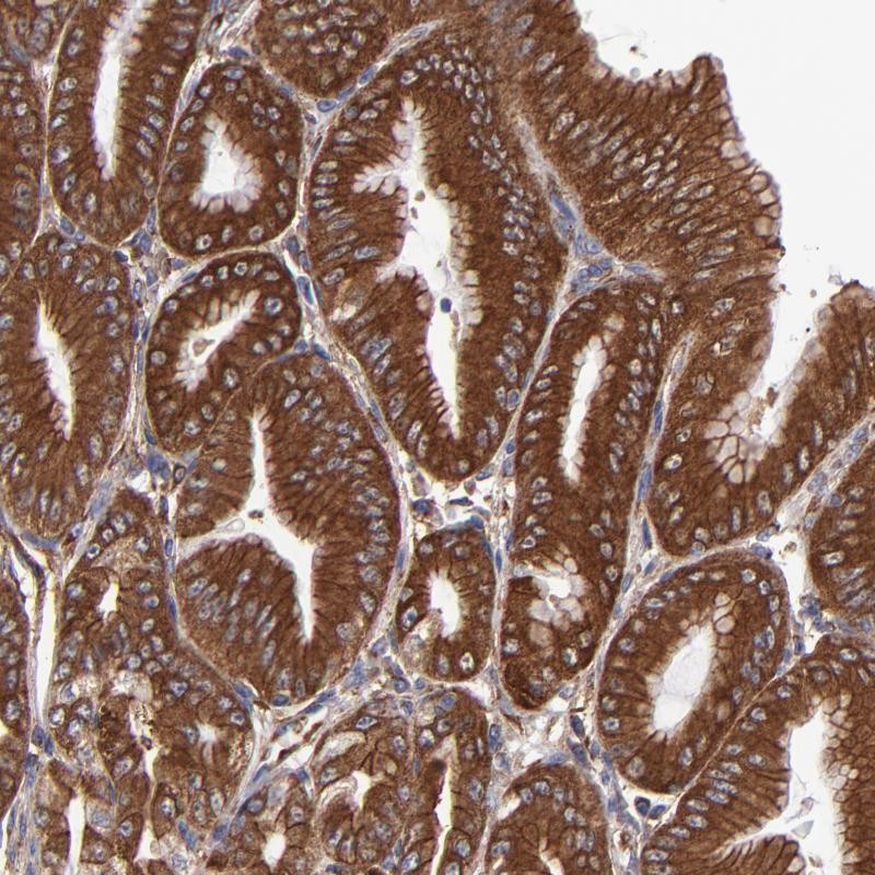
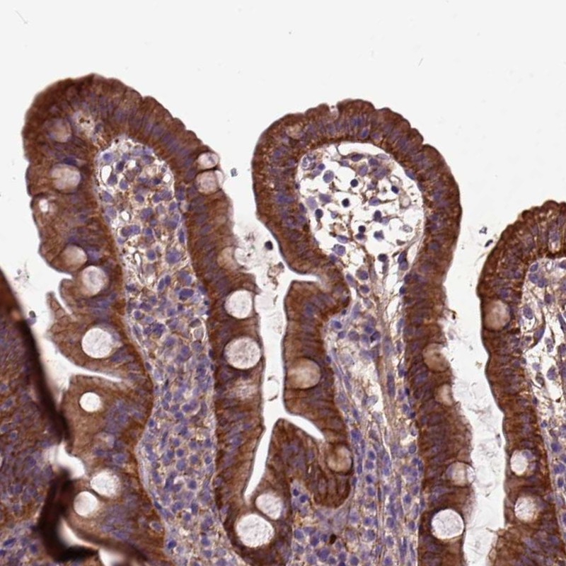
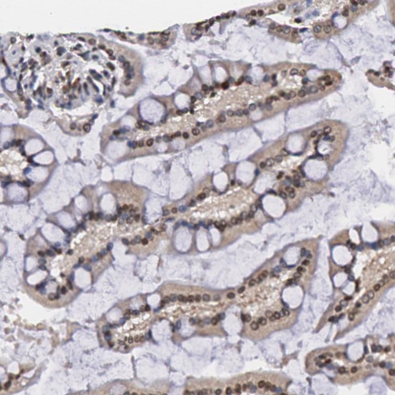
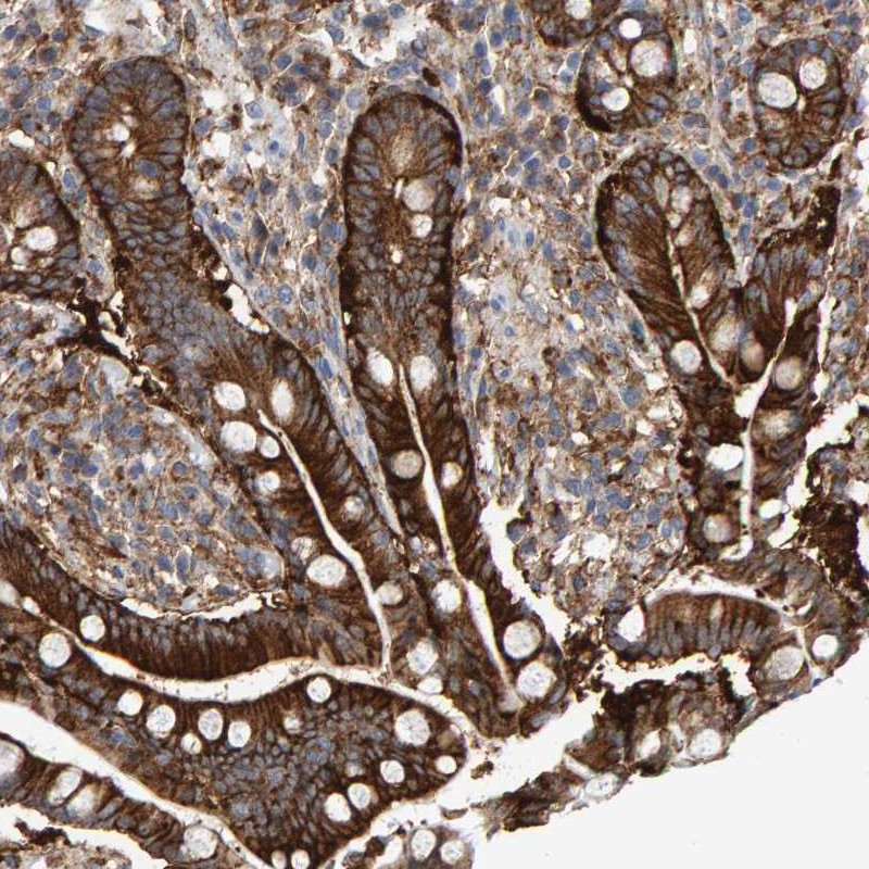
TISSUE
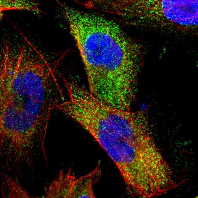
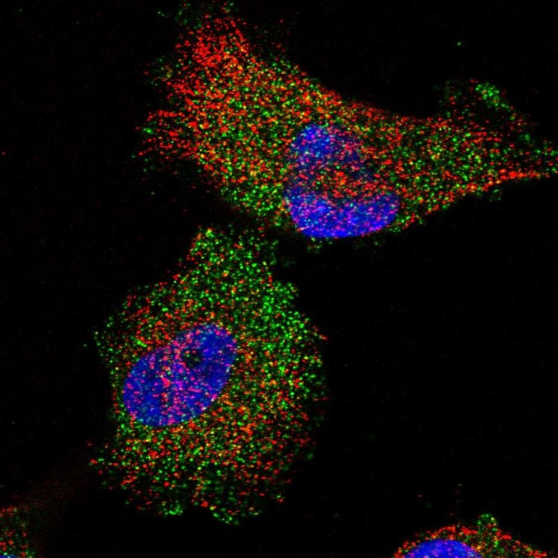
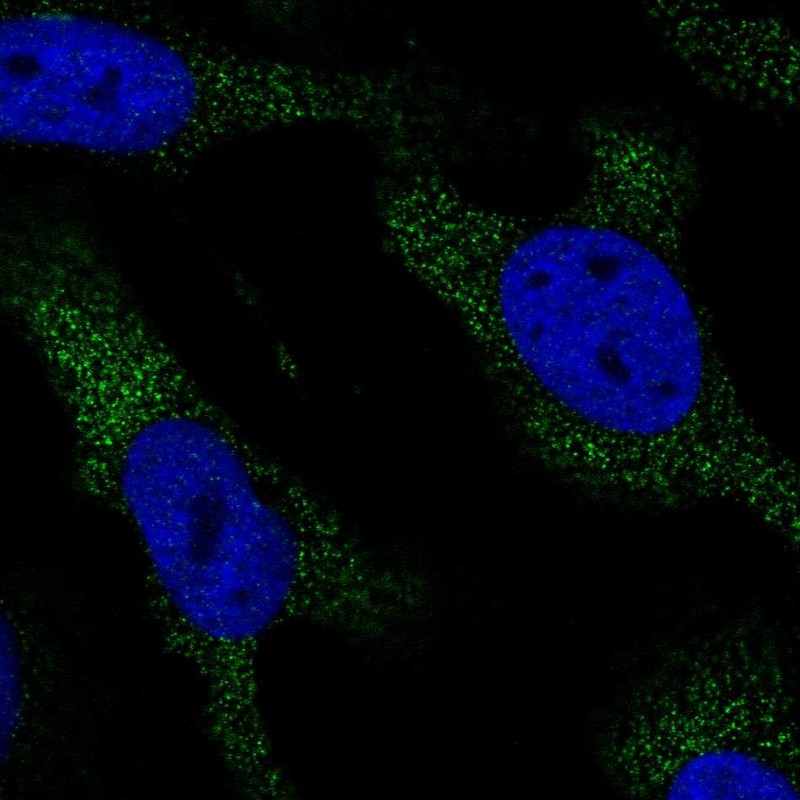
CELL
PATHOLOGY
ANTIBODY INFORMATION
Antibody HPA002321
Antibody HPA064740
Antibody CAB002752
Antibody CAB022717
Provider
Product name
Host species
Clonality
Purity
Other gene match
Released in version
References
Proper citation
VALIDATION SUMMARY
IMMUNOCYTOCHEMISTRY
Standard validation
Figure description
Antibody dilution
Literature conformity
IMMUNOHISTOCHEMISTRY
Formal validation: Orthogonal
Formal validation: Independent
Expression
Retrieval
RNA consistency
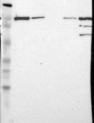
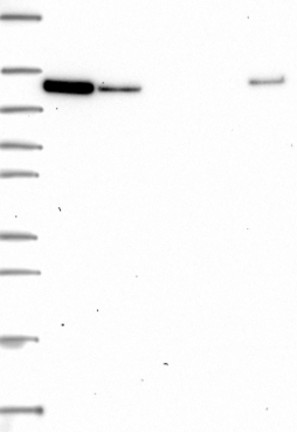
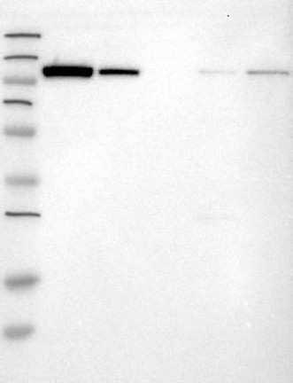
WESTERN BLOT
Target mass (kDa)
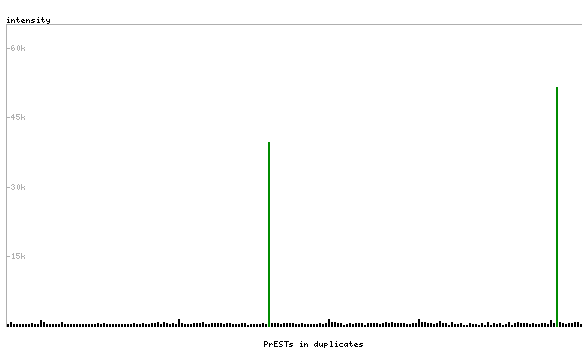
PROTEIN ARRAY
ANTIGEN INFORMATION
Antigen
Length (aa)
Antigen sequence
VFEEVLDLVDAVILTEKTALHLRARRNFRDFRGVSRRTGEEWLVTVQDTE
AHVPDVHEEVLGVVPITTLGPHNYCVILDPVGPDGKNQLGQKRVVKGEKS
FFLQPGEQLEQGIQDVYVLS
KVSHQAGDHWLIRGPLEYVPSAKVEVVEERQAIPLDENEGIYVQDVKTGK
VRAVIGSTYMLTQDEVLWEKELPPGVEELLNKGQDPLAD
Matching transcripts