TISSUE
CELL
PATHOLOGY
ANTIBODY INFORMATION
Antibody HPA006641
Antibody CAB004005
Antibody CAB044060
Antibody CAB062563
Antibody CAB068232
Provider
Product name
Host species
Clonality
Purity
Other gene match
Released in version
References
Proper citation
VALIDATION SUMMARY
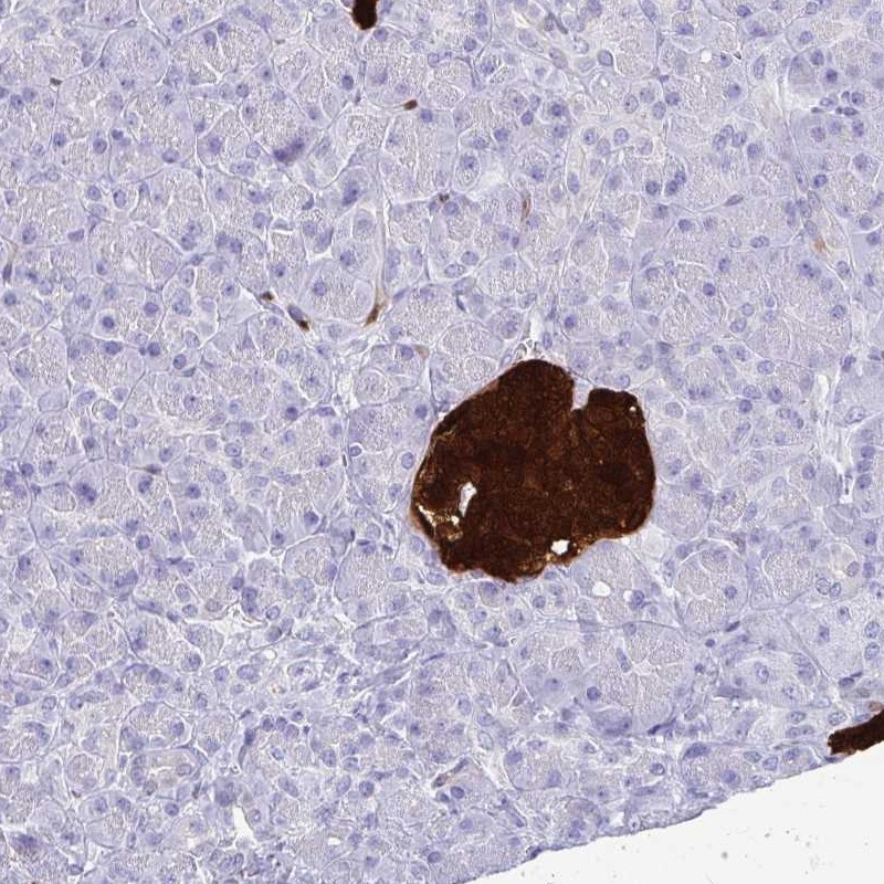
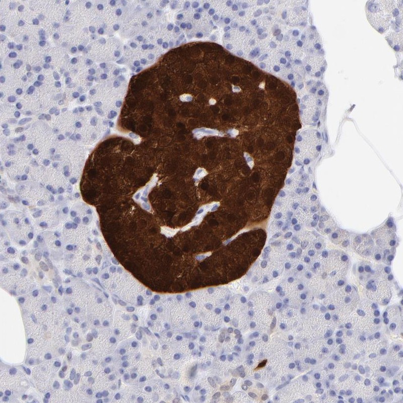
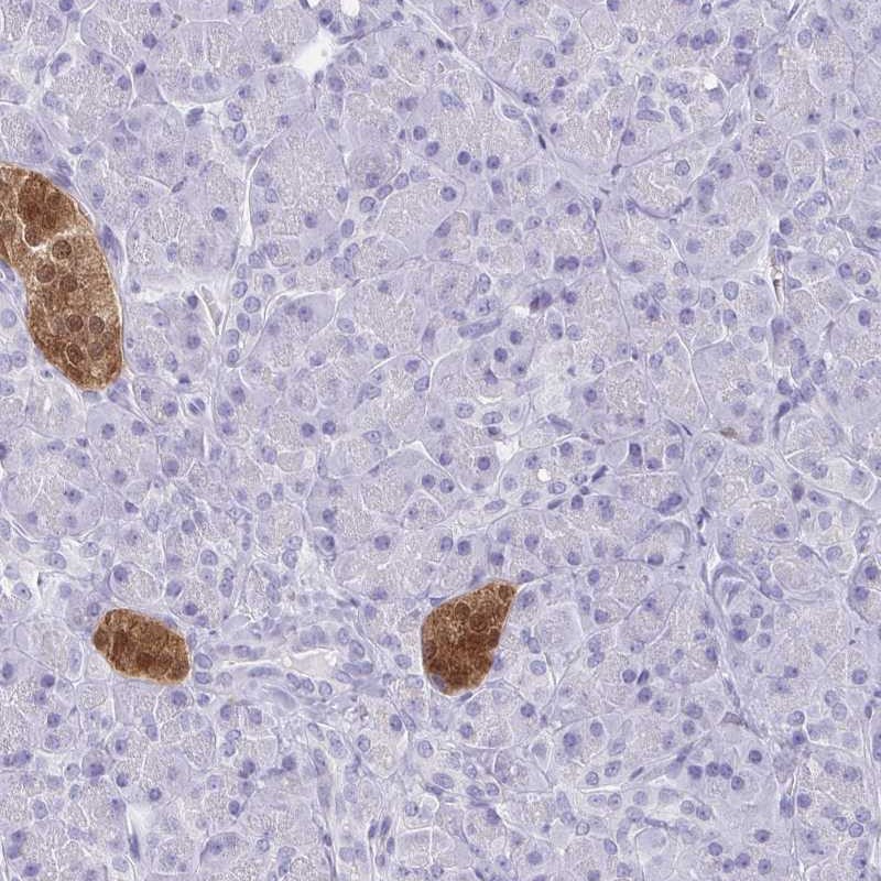
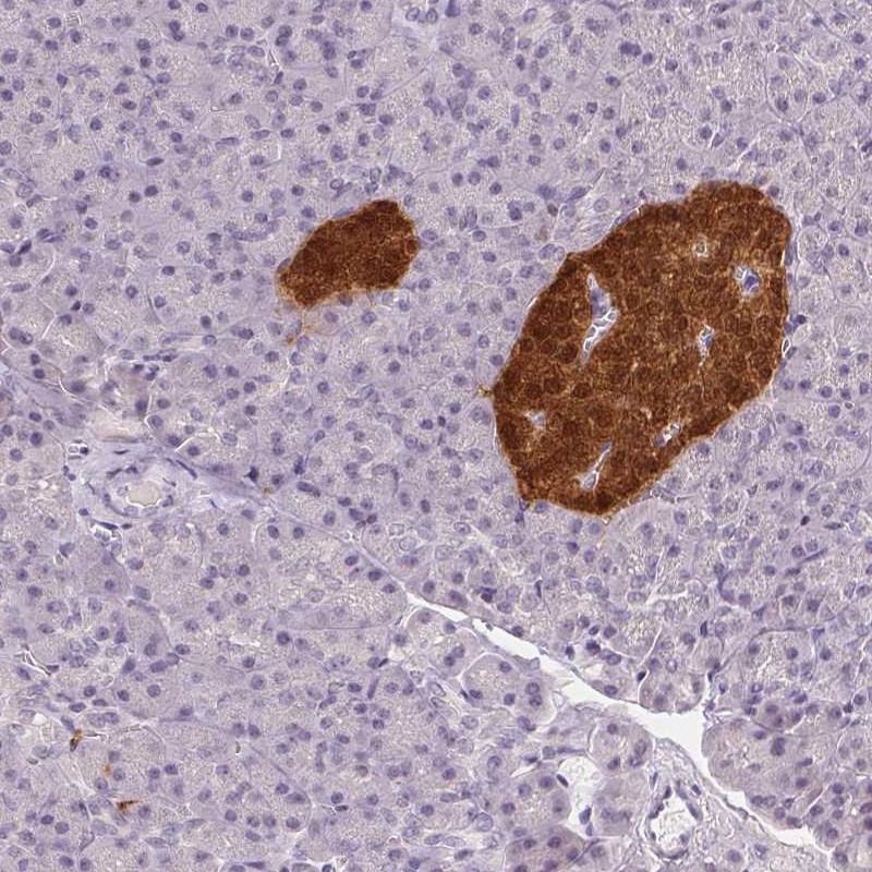
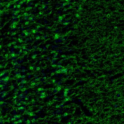
IMMUNOHISTOCHEMISTRY
Formal validation: Orthogonal
Figure description
Standard validation
Expression
Retrieval
Antibody dilution
Literature conformity
RNA consistency
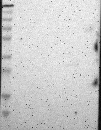
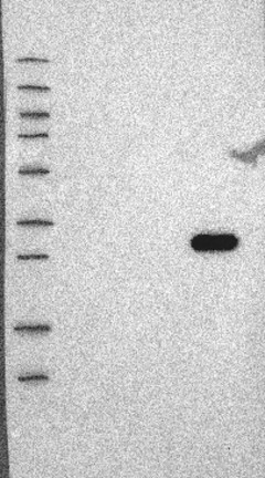
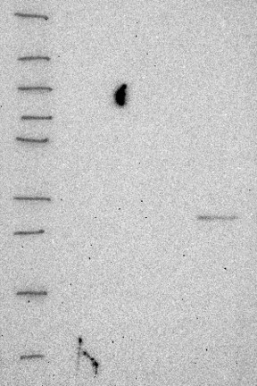
WESTERN BLOT
Target mass (kDa)
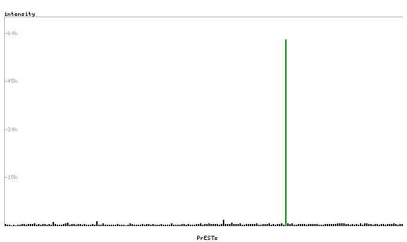
PROTEIN ARRAY
ANTIGEN INFORMATION
Antigen
Length (aa)
Antigen sequence
RDLFLHHKKAISEAKLEEYTGTMMKIFDRNKDGRLDLNDLARILALQENF
LLQFKMDACSTEERKRDFEKIFAYYDVSKTGALEGPEVDGFVKDMMELVQ
PSISGVDLDKFREILLRHCDVNKDGKIQKSELALCLGLK
Matching transcripts
RELEVANT PUBLICATIONS
1.
2.
3.
4.
5.
6.
7.
8.