TISSUE
CELL
PATHOLOGY
ANTIBODY INFORMATION
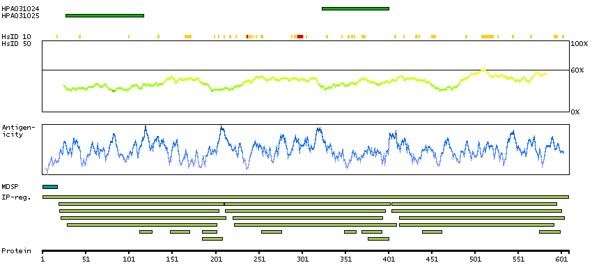
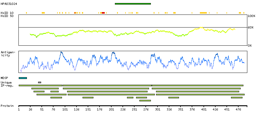
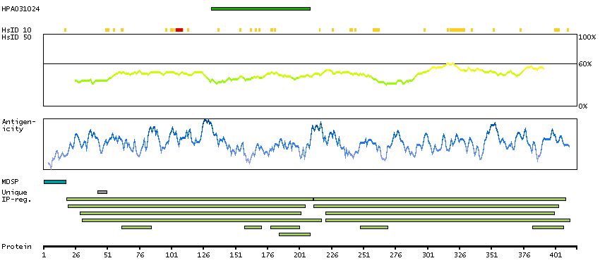
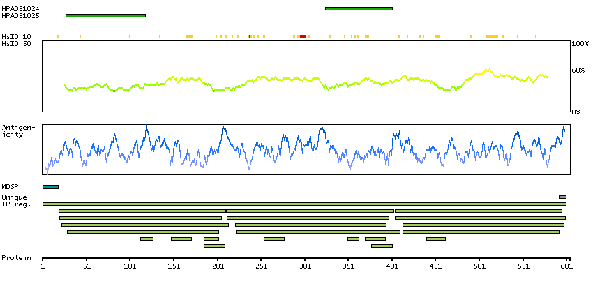
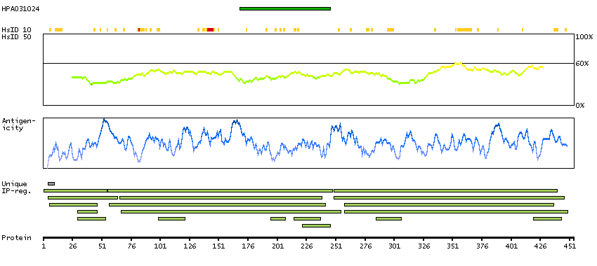
Antibody HPA031024
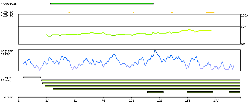
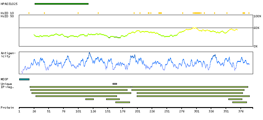
Antibody HPA031025
Antibody CAB006262
Provider
Product name
Host species
Clonality
Purity
Other gene match
Released in version
References
Proper citation
VALIDATION SUMMARY
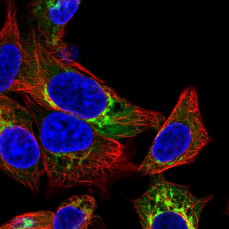
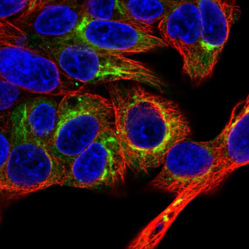
IMMUNOCYTOCHEMISTRY
Formal validation: Independent
Standard validation
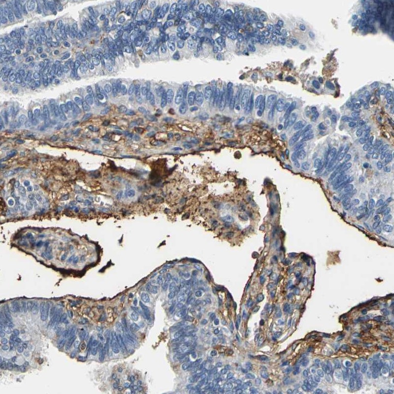
Figure description
Antibody dilution
Literature conformity
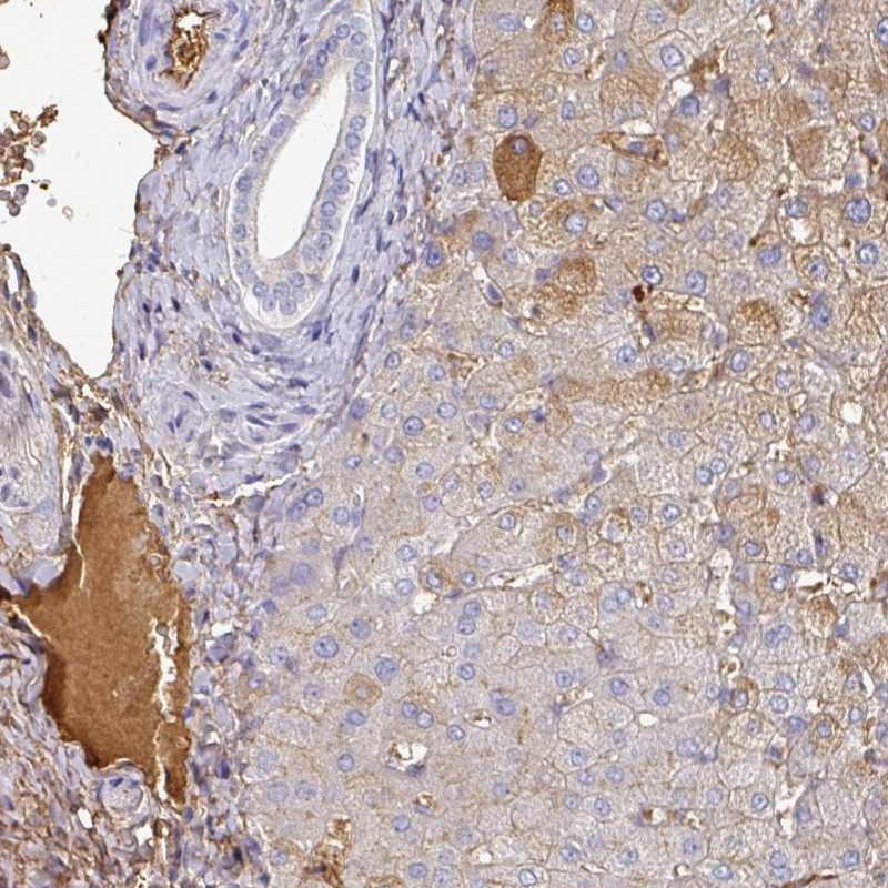
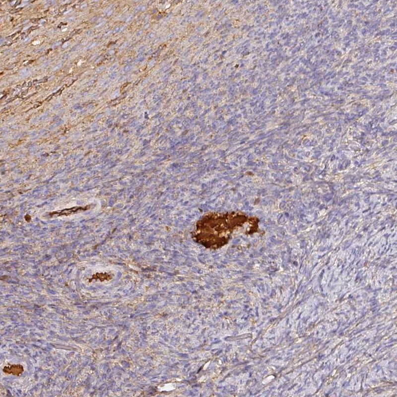
IMMUNOHISTOCHEMISTRY
Expression
Retrieval
RNA consistency
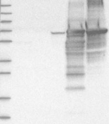
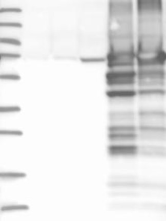
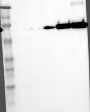
WESTERN BLOT
Target mass (kDa)
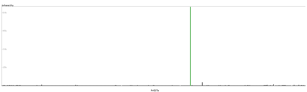
PROTEIN ARRAY
ANTIGEN INFORMATION
Antigen
Length (aa)
Antigen sequence
ADLPSLAADFVESKDVCKNYAEAKDVFLGMFLYEYARRHPDYSVVLLLRL
AKTYETTLEKCCAAADPHECYAKVFDEF
HKSEVAHRFKDLGEENFKALVLIAFAQYLQQCPFEDHVKLVNEVTEFAKT
CVADESAENCDKSLHTLFGDKLCTVATLRETYGEMADCCAKQ
Matching transcripts