TISSUE
CELL
PATHOLOGY
ANTIBODY INFORMATION
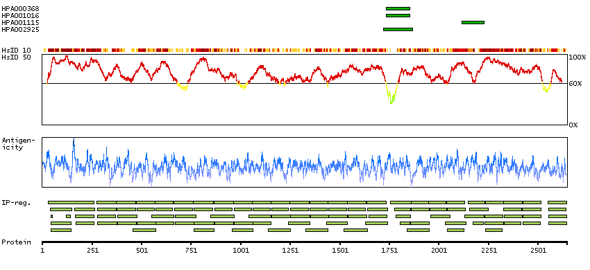
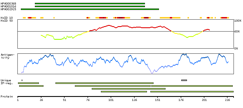
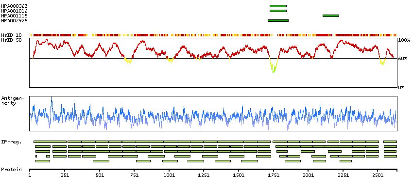
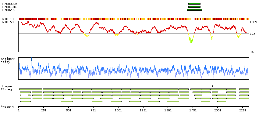
Antibody HPA000368
Antibody HPA001016
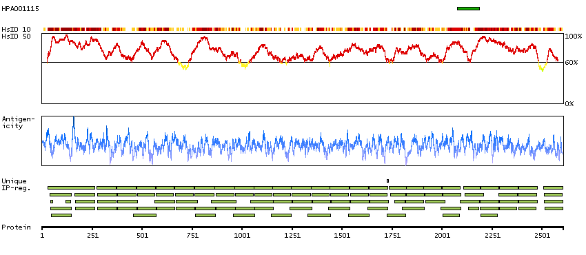
Antibody HPA001115
Antibody HPA002925
Antibody CAB000356
Provider
Product name
Host species
Clonality
Purity
Other gene match
Released in version
References
Proper citation
VALIDATION SUMMARY
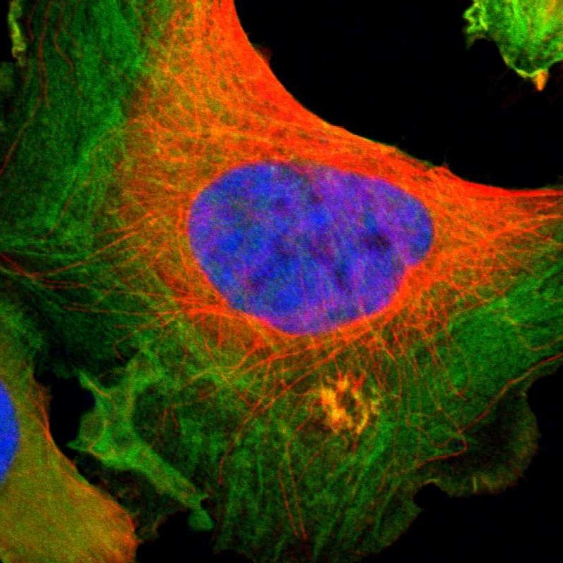
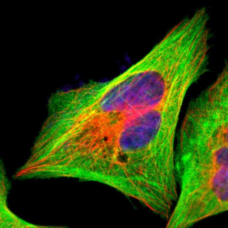
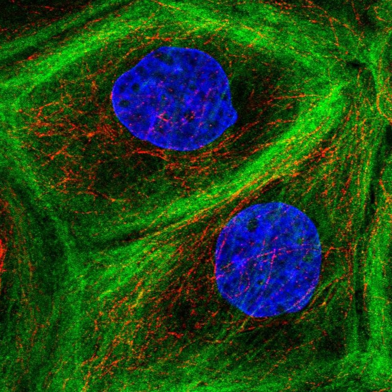
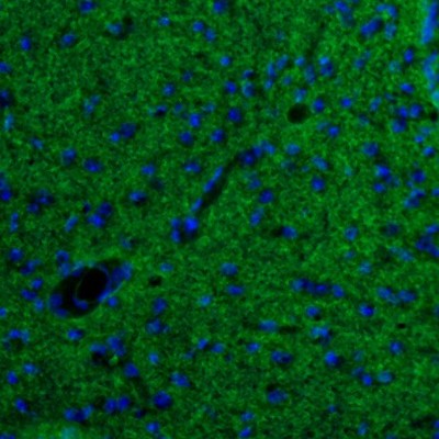
IMMUNOCYTOCHEMISTRY
Formal validation: Independent
Standard validation
Figure description
Antibody dilution
Literature conformity
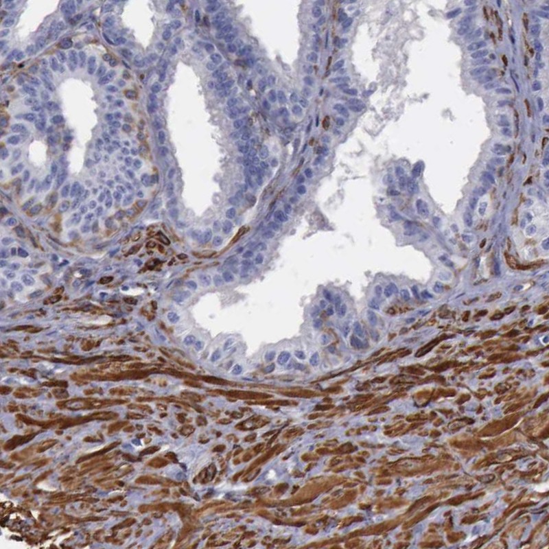
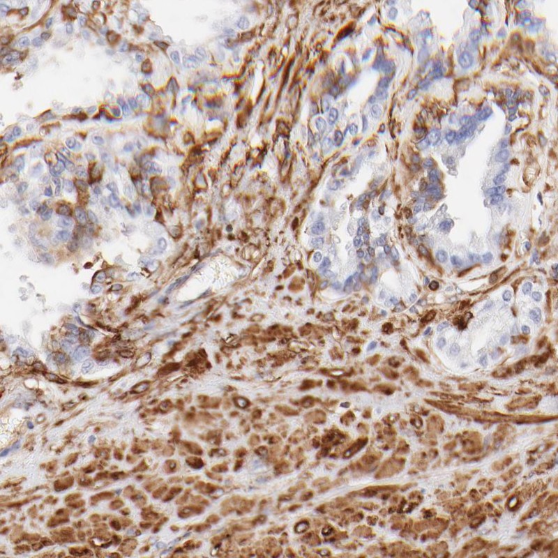
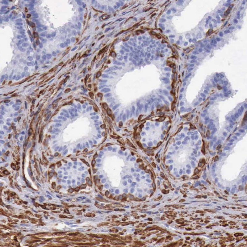
IMMUNOHISTOCHEMISTRY
Formal validation: Orthogonal
Expression
Retrieval
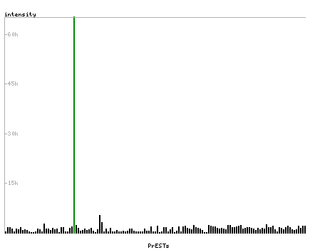
RNA consistency
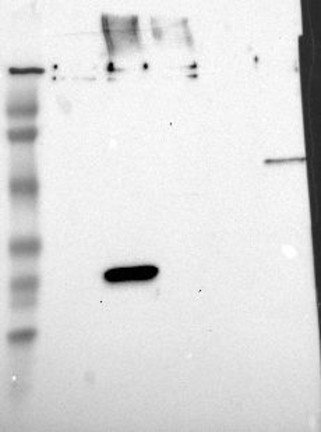
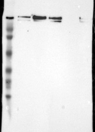
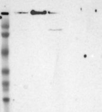
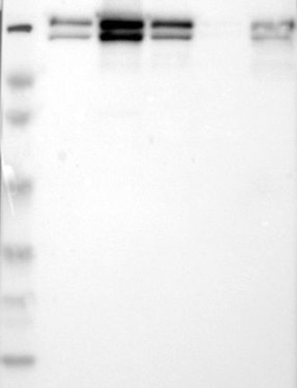
WESTERN BLOT
Target mass (kDa)
PROTEIN ARRAY
ANTIGEN INFORMATION
Antigen
Length (aa)
Antigen sequence
FQVTALAGDQPSVQPPLRSQQLAPQYTYAQGGQQTWAPERPLVGVNGLDV
TSLRPFDLVIPFTIKKGEITGEVRMPSGKVAQPTITDNKDGTVTVRYAPS
EAGLHEMDIRYDNMHIPGS
IKFADQHVPGSPFSVKVTGEGRVKESITRRRRAPSVANVGSHCDLSLKIP
EISIQDMTAQVTSPSGKTHEAEIVEGENHTYCIRFVPAEMGTHTVSVKYK
GQHVPGSPFQFTV
VICVRFGGEHVPNSPFQVTALAGDQPSVQPPLRSQQLAPQYTYAQGGQQT
WAPERPLVGVNGLDVTSLRPFDLVIPFTIKKGEITGEVRMPSGKVAQPTI
TDNKDGTVTVRYAPSEAGLHEMDIRYDNMHIPGSPLQFYVDYVNCGHVT
Matching transcripts